Figures & data
Figure 1. Effect of whole body hyperthermia on plasma glucose and insulin in db/db mice. Six-week-old db/db and db/+ mice were given free access to food and water. Whole body hyperthermia (38°C of rectal temperature for 30 min) was performed three times a week. In all panels, values are the means ± SEM of five mice, squares and solid lines represent the untreated db/db group, diamonds and solid lines represent the WBH-treated group, and solid circles and solid lines represent db/+ mice without whole body hyperthermia. (a) Fasting blood glucose concentrations in 6-week-old mice and 18-week-old mice. *p < 0.01 vs. untreated mice. (b) Fasting insulin concentrations in 10-week-old mice and 18-week-old mice. *p < 0.0001 vs. db/+ mice, #p < 0.001 vs. 10-week-old untreated mice, πp < 0.001 vs. 10-week-old untreated mice.
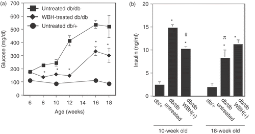
Figure 2. Effect of whole body hyperthermia on glucose and insulin tolerance tests in db/db mice. (a) For glucose tolerance tests, glucose (1.0 g/kg) was administered by intraperitoneal injection after an overnight fast. After administration of glucose, the plasma level of glucose was measured immediately before and 30, 60 120 min after administration of glucose. If insulin response against for administered glucose was appropriate, the plasma level of glucose, which was transiently increased, showed a speedy recovery. *p < 0.0001 vs. 0 min of untreated db/db mice, #p < 0.05, ##p < 0.0001 vs. 0 min of WBH-treated db/db mice. Pp < 0.05 vs. 0 min of untreated db/+ mice. (b) For insulin tolerance tests, an intraperitoneal injection of insulin (3.0 units/kg) was given after an overnight fast. After administration of insulin, the plasma level of glucose was measured immediately before and 30, 60, and 90 min after administration of insulin. If insulin sensitivity was kept appropriately, the plasma level of glucose decreased in response to administered insulin. Tail bleed samples were taken at 0, 30, 60, and 90 min for measurement of blood glucose concentrations. *p < 0.05, **p < 0.01 vs. 0 min of WBH-treated db/db mice.
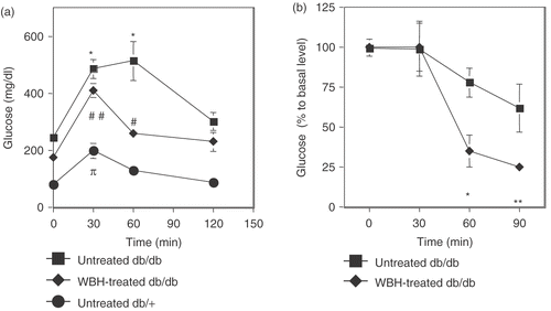
Figure 3. Effect of whole body hyperthermia on the concentrations of plasma triglyceride and free fatty acid in db/db mice. Triglyceride (a) and free fatty acid (b) concentrations were measured from fasting db/db or db/+ mice. *p < 0.05 vs. db/+ mice.
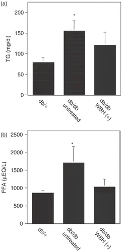
Figure 4. Effect of whole body hyperthermia on the induction of Diabetic nephropathy in db/db mice. (a) Effect of whole body hyperthermia on the urinary albumin excretion rate of db/db mice. A 24-h urine sample from each mouse was collected in metabolic cages 10 weeks after the start of this experiment. *p < 0.001 vs. db/+ mice and #p < 0.01 vs. untreated db/db mice. (b) Effect of whole body hyperthermia on accelerated mesangial expansion in db/db mice. Paraffin-embedded sections of the renal cortex were stained with periodic acid-Schiff stain. Representative light micrographs (magnification: 100 ×) from each of the groups are shown. (a) Normal glomerulus from a non-diabetic db/+ mouse at 18 weeks of age. (b) Glomerulus from an untreated db/db mouse at 18 weeks of age, showing hypertrophy and mesangial matrix expansion (arrows). (c) Glomerulus from a db/db mouse treated with 12 weeks of whole body hyperthermia until 18 weeks of age, depicting partial reversal of mesangial matrix expansion.
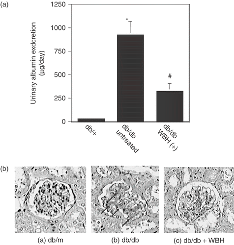
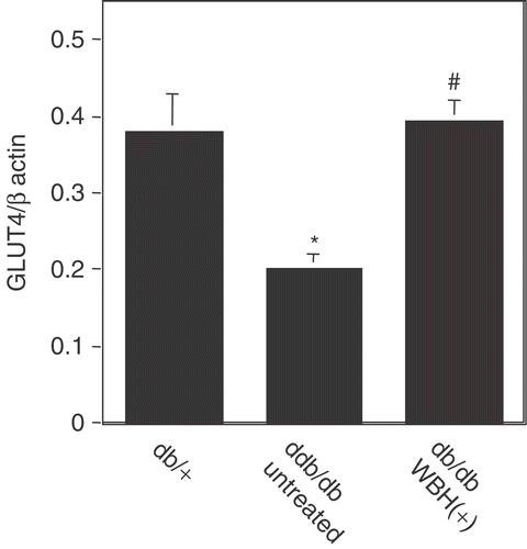
Figure 5. Real-time polymerase chain reaction analysis of GLUT4 mRNA expression in mouse skeletal muscle with or without whole body hyperthermia Total RNA was extracted from db/db or db/+ mice treated with or without whole body hyperthermia for 12 weeks and mRNA expression was analyzed by real-time polymerase chain reaction amplification. Actin was used as a control. Values are the means ± SEM of 4 mice. *p < 0.01 vs. db/+ mice and #p < 0.01 vs. untreated db/db mice.