Figures & data
Figure 1. Ultrasound guided focused ultrasound treatment of brain tumors as described in Citation[22]. Top, Left: A diagram of the treatment setting showing the skull window through which the beam is propagating into the tumor. Right: A foam mold made for each of the patients to allow positioning of the head. The mold has a hole through which the ultrasound is propagating in to the brain. Bottom, Left: A CT image of a patient in a treatment position in the head mold. The image shows a thermocouple probe that was inserted to monitor and guide the treatments. In this case the prior surgery had removed most of the tumor (shown as a fluid filled cavity with tumor in the enhancing rim). Right: An ultrasound image of a patient during the treatment showing a thermocouple probe and the tumor.
![Figure 1. Ultrasound guided focused ultrasound treatment of brain tumors as described in Citation[22]. Top, Left: A diagram of the treatment setting showing the skull window through which the beam is propagating into the tumor. Right: A foam mold made for each of the patients to allow positioning of the head. The mold has a hole through which the ultrasound is propagating in to the brain. Bottom, Left: A CT image of a patient in a treatment position in the head mold. The image shows a thermocouple probe that was inserted to monitor and guide the treatments. In this case the prior surgery had removed most of the tumor (shown as a fluid filled cavity with tumor in the enhancing rim). Right: An ultrasound image of a patient during the treatment showing a thermocouple probe and the tumor.](/cms/asset/51c5f2c7-0e14-44d7-baf4-b0c4f5ea6296/ihyt_a_219931_f0001_b.gif)
Figure 2. MRI slices, showing (left, center) the ultrasound transducer coupled via a water interface to a Rhesus monkey that has undergone a craniotomy before replacement of the skin, and an ultrasound-induced lesion (right) is enhanced following sonication (graphic courtesy of N. McDannold).
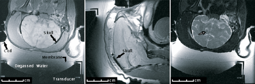
Figure 3. Top, Left: A diagram showing the wave distortion induced by a skull. Top, Right: The measured focused ultrasound field after it propagated through an ex vivo human skull showing the multiple foci induced. Bottom, Left: A diagram showing the adjustment of the phase of the array elements to compensate for the skull induced wave distortion. Bottom, Right: The measured ultrasound field after propagating through the ex vivo human skull when a CT correction algorithm was used to correct for the distortion induced by the skull Citation[49].
![Figure 3. Top, Left: A diagram showing the wave distortion induced by a skull. Top, Right: The measured focused ultrasound field after it propagated through an ex vivo human skull showing the multiple foci induced. Bottom, Left: A diagram showing the adjustment of the phase of the array elements to compensate for the skull induced wave distortion. Bottom, Right: The measured ultrasound field after propagating through the ex vivo human skull when a CT correction algorithm was used to correct for the distortion induced by the skull Citation[49].](/cms/asset/9d5bacf7-9ef2-4c01-93c9-b034a93920d7/ihyt_a_219931_f0003_b.gif)
Figure 4. A clinical prototype brain treatment system (Exablate 3000, InSightec, Inc., Haifa, Israel). (a) An illustration of the treatment planning showing how each of the ultrasound beams are propagated through a section of a skull based on the CT images obtained before the treatment and co-registered with the online MR image. (b) A photograph of the 512-element array and the mechanical positioning system. (c) A block diagram of the complete system. (d) A temperature elevation image derived from the MRI thermometry information at the end of a sonication of a monkey Citation[67]. The hotspot and the skull heating are visible in the image.
![Figure 4. A clinical prototype brain treatment system (Exablate 3000, InSightec, Inc., Haifa, Israel). (a) An illustration of the treatment planning showing how each of the ultrasound beams are propagated through a section of a skull based on the CT images obtained before the treatment and co-registered with the online MR image. (b) A photograph of the 512-element array and the mechanical positioning system. (c) A block diagram of the complete system. (d) A temperature elevation image derived from the MRI thermometry information at the end of a sonication of a monkey Citation[67]. The hotspot and the skull heating are visible in the image.](/cms/asset/0d8639d0-dfce-4dd6-9fd5-a80d2efccff4/ihyt_a_219931_f0004_b.gif)
Figure 5. Bubble-enhanced tissue destruction as seen with MRI contrast enhanced imaging after the sonication. The image shows ultrasound being delivered through an ex vivo human calvarium and brain-mimicking phantom and into a rabbit brain in vivo.
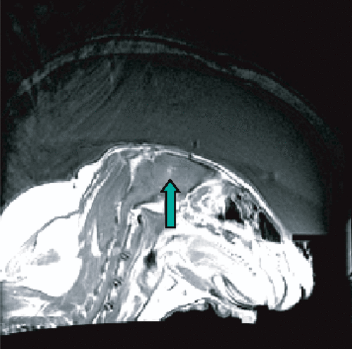
Figure 6. A T1-weighted contrast enhanced image of a rabbit brain showing two locations with disrupted blood–brain barrier Citation[80].
![Figure 6. A T1-weighted contrast enhanced image of a rabbit brain showing two locations with disrupted blood–brain barrier Citation[80].](/cms/asset/e8fce9c6-a5e6-4f94-8656-fb3108d1cff5/ihyt_a_219931_f0006_b.gif)