Figures & data
Figure 1. Determination of the member of Hsp70 family detected by clone W27. Exponentially growing NHD cells were heat shocked at 43°C for 2 h, and the proteins were extracted after incubation at 37°C for indicated times. (a) The same blot was probed with clone W27 and clone C92F3A-5. (b) The upper blot was probed with clone 1B5, while the lower blot was probed with clone W27. (c) The same blot was separated into two parts, and both bands were probed with clone N27F3-4, while only the lower band was probed with clone W27.
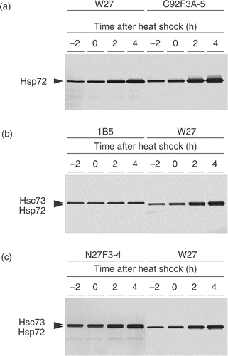
Figure 2. Time-course experiment of Hsp72 induction in NHD cells after heat shock. Exponentially growing NHD cells were treated at 43°C for 2 h. (a) Typical Western blotting pattern of Hsp72 induction detected by clone W27. (b) The relative amount of Hsp72 measured by Western blotting (•) and LSC (○); the relative amount was expressed as the ratio of the amount of Hsp72 to that of unheated NHD cells. The band intensity of the blots was determined by densitometrical analysis.
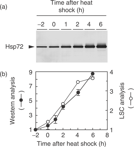
Figure 3. Augmented expression of Hsp72 protein in unheated human tumour cell lines. (a) Total proteins were extracted from exponentially growing cells, and the Hsp72 level was examined by Western blotting using clone W27. (b) HT1080 cells grown on cover-slips were fixed with methanol and stained with clone W27, followed by the incubation with FITC-conjugated anti-mouse Ig. Hsp72 is depicted in white. (c) Relationship between the relative intracellular amount of Hsp72 determined by Western blotting and the relative nuclear expression of Hsp72 measured by LSC. A close correlation was observed between them (R = 0.906).

Figure 4. Relationship between the colony-forming ability in soft agar and relative amount of Hsp72 determined by Western blotting and LSC. (a) The nuclear expression of Hsp72 was measured by LSC. The colony-forming ability was determined in 0.3% soft agar as described in the Materials and methods. A close correlation was observed between them (R = 0.880). (b) The total amount of Hsp72 was measured by Western blotting. The colony-forming ability was determined as described above. A close correlation was observed between them (R = 0.881).
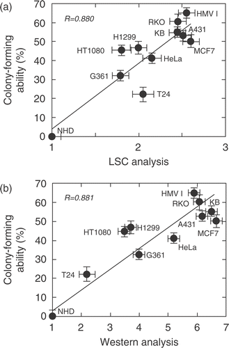
Table I. Augmented expression of Hsp72 in breast cancer tissues.