Figures & data
Figure 1. (a) Schematic illustration of the dual mirror microwave technique for thermal therapy of localized breast cancer (not drawn to scale). (b) Arrangement of the mirror and breast coronal section in the computational model; a1 = a2 = 1.25λc, b1 = b2 = 3.75λc, c1 = c2 = D, d1 = d2 = 2λc, D = max{Xlen, Ylen}; s1, s2: sources (figure drawn approximately to scale; see for λc).
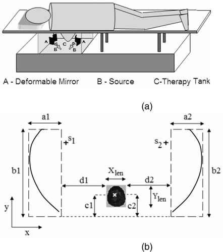
Figure 2. (a) Focusing strategy (εr2: tissue dielectric permittivity; εr1: couplant dielectric permittivity; μ0: magnetic permeability), and (b) surface tangents inside S for adaptive field focusing.
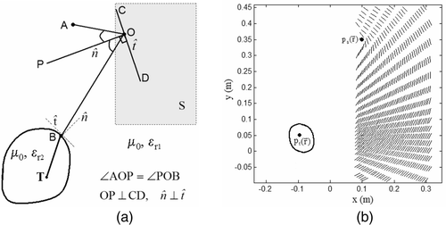
Figure 3. Mirror shape estimation (a) Mirror deformations estimated for given source and tumor locations. (b) Path lengths from to
computed for mirror 1 in (3a).
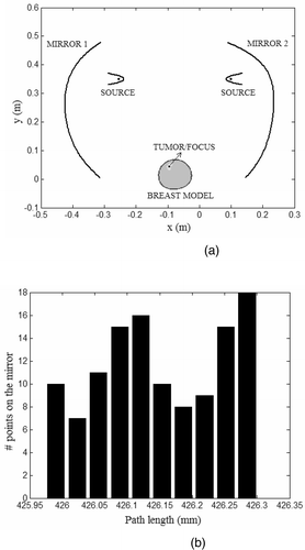
Figure 4. Two-dimensional representation of the (a) dual mirror microwave technique for hyperthermia, (b) computational model.
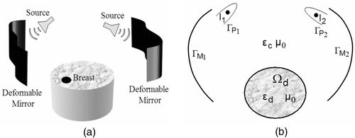
Figure 5. Normalized field magnitude maintained inside the tank. (a) 500 MHz and (b) 700 MHz for single mirror; (c) 500 MHz and (d) 700 MHz for dual mirror (x denotes target location).
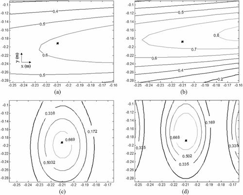
Figure 6. Tissue permittivity map for the MR breast image. Boundary extraction for (a) skin and (b) tumor; (c) relative permittivity and (d) electrical conductivity (S/m) obtained using Equation 10 and Citation[10], Citation[24].
![Figure 6. Tissue permittivity map for the MR breast image. Boundary extraction for (a) skin and (b) tumor; (c) relative permittivity and (d) electrical conductivity (S/m) obtained using Equation 10 and Citation[10], Citation[24].](/cms/asset/94300477-d3d5-464c-a229-b8a7b42ad9f8/ihyt_a_272665_f0006_b.gif)
Table I. Patient information for MR derived breast models.
Figure 7. Coronal MR breast images used in the hyperthermia simulations (a) patient data I, (b) patient data II, (c) patient data III, (d) patient data IV (black contours indicate tumor sites).
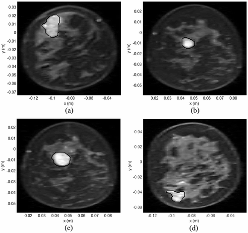
Figure 8. Tissue SAR (W/kg/m) inside MR derived breast models for which and
(a) Model I, (b) Model II, (c) Model III, (d) Model IV.
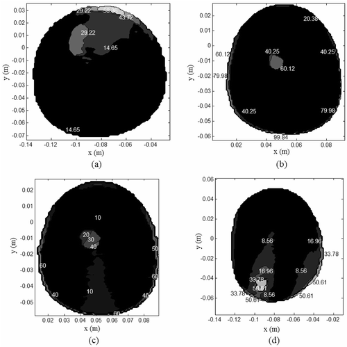
Table II. Electrical and thermal properties of breast tissues Citation[10], Citation[24–26] (ε = ε′–jε″, couplant permittivity, εc = 43.76-j22.82, cb = 4000 (J/kg/°C), Tb = 37°C, f = 500 MHz, λc = 0.0907m).
Figure 9. Steady state temperature for the tissue SAR in (T0 = 30°C), (a) Model I, (b) Model II, (c) Model III, (d) Model IV.
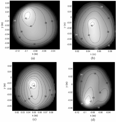
Table III. Tumor and normal tissue damage for the hyperthermia simulations.
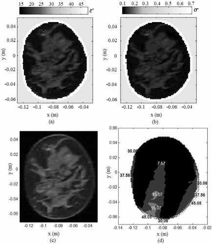
Figure 10. Hyperthermia simulation for model III with 60% decrease in εtumor. (a) Dielectric constant; (b) electrical conductivity at 500 MHz using Equation 10 and Citation[10], Citation[24]; (c) steady state thermal contours.
![Figure 10. Hyperthermia simulation for model III with 60% decrease in εtumor. (a) Dielectric constant; (b) electrical conductivity at 500 MHz using Equation 10 and Citation[10], Citation[24]; (c) steady state thermal contours.](/cms/asset/ece649b4-22a3-4db0-b754-54af33238c2d/ihyt_a_272665_f0010_b.gif)
Figure 11. EM energy deposited inside the benign breast tissue for field focus at (−0.093, − 0.0475). (a) Tissue dielectric constant, (b) electrical conductivity, (c) coronal MR breast image, (d) Tissue SAR within the breast model (W/kg/m).