Figures & data
Figure 1. The effect of hyperthermia on the viability (A and B) and growth (C and D) of K562 cells at various times after heat treatment. Cells were heat treated at 43°C (A and C) and 45°C (B and D) for different periods of time. The number of cells and their viability was determined 24, 72 and 120 h of incubation at 37°C after heat treatment. Values are Mean ± SEM from three independent experiments.
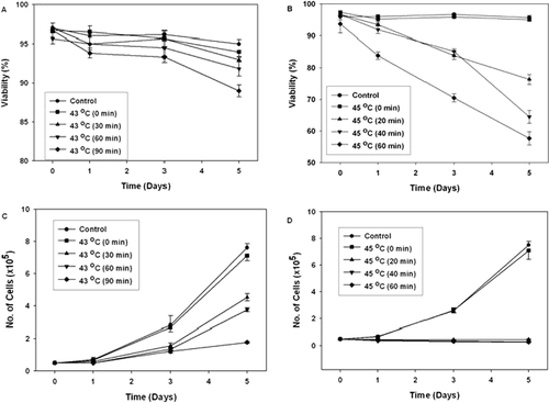
Figure 2. The effect of hyperthermia on glycophorin A expression in K562 cells. Cells were heat treated for different periods of time at 43°C and incubated for 120 hr after heat treatment at 37°C. Then the cells were stained with FITC-conjugated anti glycophorin A, and the green fluorescence of the stained cells was measured by flow cytometry. (A–D) K562 cells were heat treated at 43°C for 0 min, 30 min, 60 min and 90 min, respectively. (E) As positive control for glycophorin A expression, cells were treated with sodium butyrate (1mM). (Dark Gray line: Control (37°C); Light Gray line: Heat or Sodium butyrate treatement).
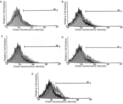
Table I. The effect of hyperthermia (43°C and 45°C) on induction of differentiation (haemoglobin synthesis and glycophorin A expression) in K562 cells. Cells were heated at these temperatures for different periods of time and incubated for 120 h after hyperthermia. As positive controls, cells were treated with sodium butyrate (SB) and hemin. The fraction of benzidine positive cells and glycophorin A-expressing cells are represented as Mean ± SEM from three independent experiments. B+ cells: Benzidine positive cells, Gly A-exp cells: Glycophorin A-expressing cells. Compared with control (37°C); *P < 0.05; ♦P < 0.01.
Table II. The effect of hyperthermia (43°C) on induction of apoptosis in K562 cells. Cells were heated at 43°C for different periods of time and studied at 24, 72 and 120 h after heat treatment. Annexin-V and PI staining were measured by flow cytometry. The results of quantitative analysis of Annexin-V/PI staining are represented as mean ± SEM from three independent experiments.
Figure 3. The effect of hyperthermia on Annexin-V binding/PI uptake in K562. Cells were heated at different periods of time at 45°C and incubated for 24 h (A), 72 h (B) and 120 h (C) after heat treatment. Annexin-V and PI staining were measured by flow cytometry. The dual fluorescence dot plots show the normal (viable) cells in the lower left quadrant (Annexin−/PI−), the early apoptotic cells in the lower right quadrant (Annexin+/PI−) and the late apoptotic or necrotic cells in the upper right quadrant (Annexin+/PI+). Cell debris was excluded from the analysis by conventional gating of forward scatter versus side scatter dot plots. X axis: The fluorescence intensity of Annexin-V-FITC and Y axis: The fluorescence intensity of Propidium Iodide (PI).
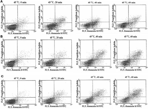
Figure 4. Morphological assessment of apoptosis and necrosis in K562 cells. Cells were heat treated at 45°C for different periods of time and studied 72 h after heat treatment. AO/EtBr double staining of K562 cells shows viable cells are uniformly green, early apoptotic cells are green with bright green dots in their nuclei (green arrows) and late apoptotic cells (orange arrows) are orange and in contrast to necrotic cells (orange arrows, N), they show condense fragmented nuclei. (A) Control K562 cells, (B-E) cells were heat treated at 45°C for 0, 20, 40 and 60 min, respectively. Cells were observed under fluorescence microscope at 400×.
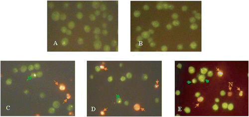
Table III. The effect of hyperthermia (45°C) on induction of apoptosis and necrosis in K562 cells. Cells were heated at 45°C for different periods of time and studied at 72 h after heat treatment. The results of quantitative analysis of AO/EtBr double staining are represented as mean ± SEM. Compared with control (37°C); *P < 0.05, ♦P < 0.01.
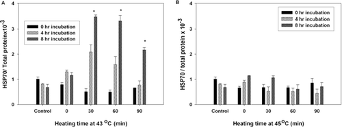
Figure 5. The effect of hyperthermia on HSP70 level in K562 cells. Cells were heat treated at 43°C (A) and 45°C (B) for different periods of time and studied 0, 4 and 8 h after heat treatment. The x-axis shows the temperature and the time of heating, the y-axis shows the level of HSP70 as determined by the ELISA HSP70 kit. The results are expressed as the ratio of HSP70 protein/total protein from two independent experiments (each performed in duplicate). P < 0.02 compared to the control samples (37°C).