Figures & data
Figure 1. Left: CBE predicted over the temperature range employed in this study using Equation 1 for single lipid and aqueous scatterers with thermal properties given in our initial theoretical study Citation[16]. Right: CBE from simulated B-mode images with collections of scatterers in the presence of noise Citation[20].
![Figure 1. Left: CBE predicted over the temperature range employed in this study using Equation 1 for single lipid and aqueous scatterers with thermal properties given in our initial theoretical study Citation[16]. Right: CBE from simulated B-mode images with collections of scatterers in the presence of noise Citation[20].](/cms/asset/c1ccd168-72ee-4640-a1c9-701eadf8006f/ihyt_a_294385_f0001_b.gif)
Figure 2. Configuration for automatic heating and ultrasonic imaging of nude mice. Deionized and degassed water was heated to equilibrium under control of the laptop-based imaging system.
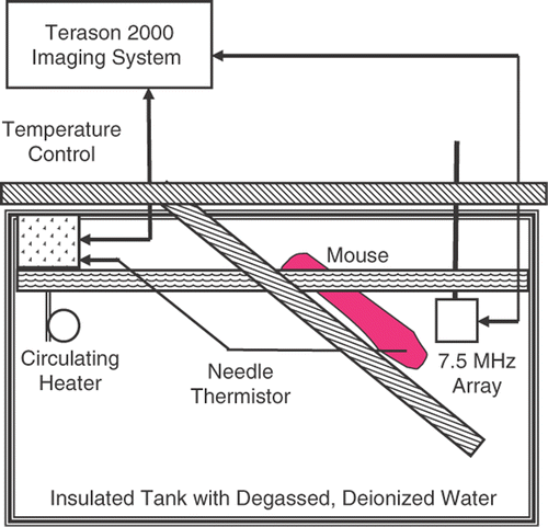
Figure 3. Small animal hyperthermia ultrasound system (SAHUS) for heterogeneous heating of tissue near the 14 mm diameter, 5 MHz transducer.
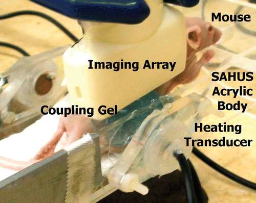
Figure 4. Ultrasonic image of a live nude mouse. Focal zone of the 7.5 MHz transducer was at 2 cm.
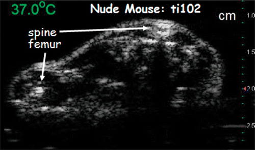
Figure 5. Image region at 37°C and 2D motion fields over 0.5°C steps at 3°C intervals during homogeneous heating estimated using optimization techniques. Image intensity is in dB. Arrow lengths are 50 times the actual motion. The magnitude of the largest motion was about 100 μm. Motion values were found via bilinear interpolation over the image.
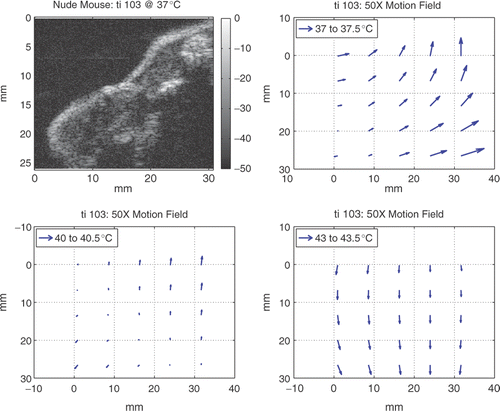
Figure 6. CBE measured over the thighs of live nude mice. Means of CBE at pixels with increasing BE and means at pixels with decreasing BE are shown from four experiments.
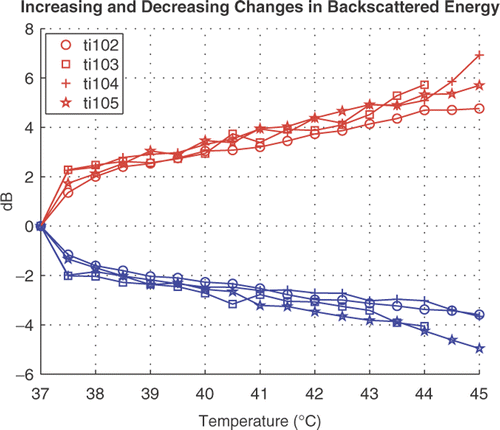
Figure 7. Upper panel: Terason 3000 image at 7.5 MHz of an HT29 tumor in the leg of a nude mouse heated to 43°C at the thermocouple. The circle shows the position and size of the heating transducer. Lower panel: CBE at 43°C compared to body temperature for the inset region above the thermocouple probe in the conventional image. Tissue volume for the CBE image is 0.075 cm3. Vertical bars show the image intensities in dB.
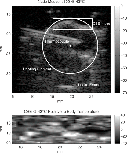
Figure 8. Mean positive CBE at 43°C relative to body temperature in a portion of implanted HT29 tumors in three nude mice after heterogeneous heating in our SAHUS apparatus shown in Citation[27].
![Figure 8. Mean positive CBE at 43°C relative to body temperature in a portion of implanted HT29 tumors in three nude mice after heterogeneous heating in our SAHUS apparatus shown in Figure 3 Citation[27].](/cms/asset/e455ca71-1f82-45d9-8937-9de61966ea1b/ihyt_a_294385_f0008_b.gif)