296
Views
0
CrossRef citations to date
0
Altmetric
Original

p53 status-dependent sensitization of human tumour cells to hyperthermia by plant flavonol

, , , , &
Pages 415-424 | Received 09 Aug 2007, Accepted 18 Mar 2008, Published online: 09 Jul 2009

Figures & data

Table I.  List of human tumour cells used in this study.

Figure 1. Effects of 0.15% DMSO on the levels of Hsp72 and p53 in human cancer cell lines. Exponentially growing cells were treated with or without 0.15% DMSO for 2 h. Whole-cell proteins were extracted and analysed with Western blotting. (a) Hsp72 and p53 were visualized using anti-Hsp72/73 antibody and anti-p53 antibody, respectively. C, control (without any treatment). (b) The intensity of the band indicated in (a) was measured by densitometry and the relative level of Hsp72 protein was calculated. The protein level detected in the control cells was determined as 1.0. Filled column, control; open column, DMSO.
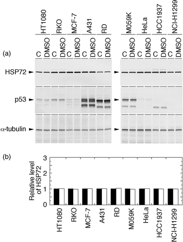
Figure 1. Effects of 0.15% DMSO on the levels of Hsp72 and p53 in human cancer cell lines. Exponentially growing cells were treated with or without 0.15% DMSO for 2 h. Whole-cell proteins were extracted and analysed with Western blotting. (a) Hsp72 and p53 were visualized using anti-Hsp72/73 antibody and anti-p53 antibody, respectively. C, control (without any treatment). (b) The intensity of the band indicated in (a) was measured by densitometry and the relative level of Hsp72 protein was calculated. The protein level detected in the control cells was determined as 1.0. Filled column, control; open column, DMSO.

Figure 2. Effects of heat shock and heat shock plus QCT on the levels of Hsp72 and p53 in human cancer cell lines. Exponentially growing cells were heat-shocked at 43°C alone for 2 h or concomitantly with QCT (150 µM). Control cells were neither heat-shocked nor treated with QCT. Whole-cell proteins were extracted immediately after heat shock and analysed with Western blotting. (a) Hsp72 and p53 were visualized using anti-Hsp72/73 antibody and anti-p53 antibody, respectively. C, control (without any treatment); HS, heat shock; H/Q, heat shock plus QCT. (b) The intensity of the band indicated in (a) was measured by densitometry and the relative level of Hsp72 protein was calculated. The protein level detected in the control cells was determined as 1.0. Filled column, control; dotted column, heat shock; open column, heat shock plus QCT.
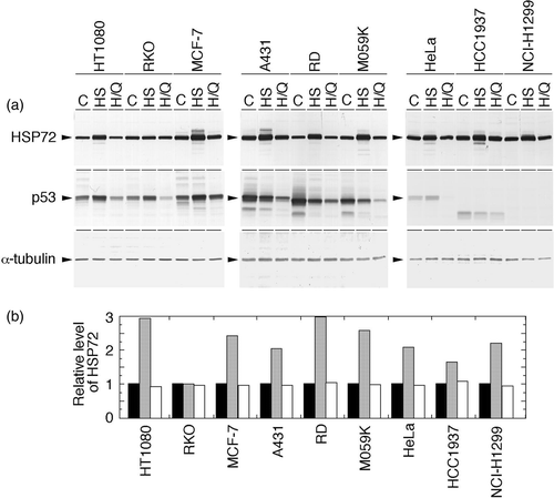
Figure 2. Effects of heat shock and heat shock plus QCT on the levels of Hsp72 and p53 in human cancer cell lines. Exponentially growing cells were heat-shocked at 43°C alone for 2 h or concomitantly with QCT (150 µM). Control cells were neither heat-shocked nor treated with QCT. Whole-cell proteins were extracted immediately after heat shock and analysed with Western blotting. (a) Hsp72 and p53 were visualized using anti-Hsp72/73 antibody and anti-p53 antibody, respectively. C, control (without any treatment); HS, heat shock; H/Q, heat shock plus QCT. (b) The intensity of the band indicated in (a) was measured by densitometry and the relative level of Hsp72 protein was calculated. The protein level detected in the control cells was determined as 1.0. Filled column, control; dotted column, heat shock; open column, heat shock plus QCT.

Figure 3. Effects of heat shock and heat shock plus QCT on nuclear Hsp72 levels in human cancer cell lines. Exponentially growing cells were heat-shocked at 43°C alone for 2 h or concomitantly with QCT (150 µM). Control cells were neither heat-shocked nor treated with QCT. Nuclear proteins were extracted immediately after heat shock and analysed with Western blotting. (a) Hsp72 and p53 were visualized using anti-Hsp72/73 antibody and anti-p53 antibody, respectively. C, control (without any treatment); HS, heat shock; H/Q, heat shock plus QCT. (b) The intensity of the band indicated in (a) was measured by densitometry and the relative level of Hsp72 protein was calculated. The protein level detected in the heat-shocked cells was determined as 100%. Filled column, control; dotted column, heat shock; open column, heat shock plus QCT.
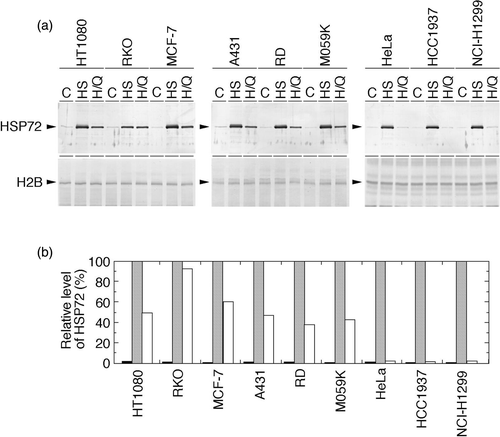
Figure 3. Effects of heat shock and heat shock plus QCT on nuclear Hsp72 levels in human cancer cell lines. Exponentially growing cells were heat-shocked at 43°C alone for 2 h or concomitantly with QCT (150 µM). Control cells were neither heat-shocked nor treated with QCT. Nuclear proteins were extracted immediately after heat shock and analysed with Western blotting. (a) Hsp72 and p53 were visualized using anti-Hsp72/73 antibody and anti-p53 antibody, respectively. C, control (without any treatment); HS, heat shock; H/Q, heat shock plus QCT. (b) The intensity of the band indicated in (a) was measured by densitometry and the relative level of Hsp72 protein was calculated. The protein level detected in the heat-shocked cells was determined as 100%. Filled column, control; dotted column, heat shock; open column, heat shock plus QCT.
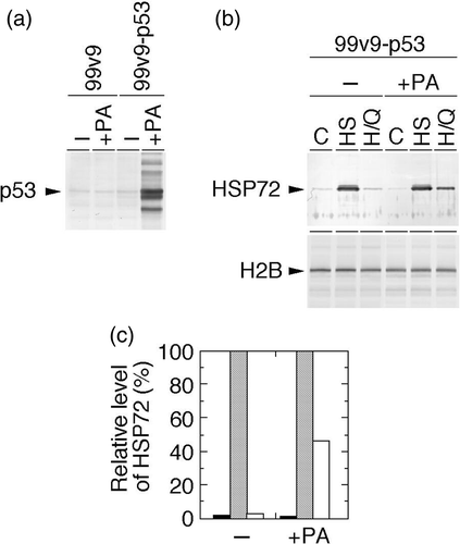
Table II.  Effects of quercetin treatment on lethal effects of heat shock.

Figure 4. Cellular p53 expression and effects of heat shock and heat shock plus QCT on nuclear Hsp72 levels in human p53-inducible cells. 99v9 cells having deleted p53 and their derivative 99v9–p53 cells having ponasterone A (PA)-inducible vectors for human wild-type p53 gene were used. (a) After treatment with or without PA for 24 h, whole-cell proteins were extracted and analysed with Western blotting. p53 was visualized using anti-p53 antibody. (b) After treatment with or without PA for 24 h, exponentially growing 99v9–p53 cells were heat-shocked at 43°C alone for 2 h or concomitantly with QCT (150 µM). Control cells were neither heat-shocked nor treated with QCT. Nuclear proteins were extracted immediately after heat shock and analysed with Western blotting. Hsp72 was visualized using anti-Hsp72/73 antibody. C, control (without any treatment); HS, heat shock; H/Q, heat shock plus QCT. (c) The intensity of the band indicated in (b) was measured by densitometry and the relative level of Hsp72 protein was calculated. The protein level detected in the heat-shocked cells was determined as 100%. Filled column, control; dotted column, heat shock; open column, heat shock plus QCT.

Figure 4. Cellular p53 expression and effects of heat shock and heat shock plus QCT on nuclear Hsp72 levels in human p53-inducible cells. 99v9 cells having deleted p53 and their derivative 99v9–p53 cells having ponasterone A (PA)-inducible vectors for human wild-type p53 gene were used. (a) After treatment with or without PA for 24 h, whole-cell proteins were extracted and analysed with Western blotting. p53 was visualized using anti-p53 antibody. (b) After treatment with or without PA for 24 h, exponentially growing 99v9–p53 cells were heat-shocked at 43°C alone for 2 h or concomitantly with QCT (150 µM). Control cells were neither heat-shocked nor treated with QCT. Nuclear proteins were extracted immediately after heat shock and analysed with Western blotting. Hsp72 was visualized using anti-Hsp72/73 antibody. C, control (without any treatment); HS, heat shock; H/Q, heat shock plus QCT. (c) The intensity of the band indicated in (b) was measured by densitometry and the relative level of Hsp72 protein was calculated. The protein level detected in the heat-shocked cells was determined as 100%. Filled column, control; dotted column, heat shock; open column, heat shock plus QCT.

Table III.  Effects of p53 induction on QCT enhancement of heat-sensitivity.

Reprints and Corporate Permissions

Please note: Selecting permissions does not provide access to the full text of the article, please see our help page How do I view content?

To request a reprint or corporate permissions for this article, please click on the relevant link below:

Academic Permissions

Please note: Selecting permissions does not provide access to the full text of the article, please see our help page How do I view content?

Obtain permissions instantly via Rightslink by clicking on the button below:

If you are unable to obtain permissions via Rightslink, please complete and submit this Permissions form. For more information, please visit our Permissions help page.