Figures & data
Figure 1. Thermotherapy treatment of the pelvic region after intratumoral injection of magnetic nanoparticles using the alternating magnetic field applicator MFH 300F® (MagForce Nanotechnologies AG, Berlin, Germany).
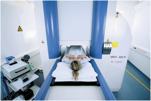
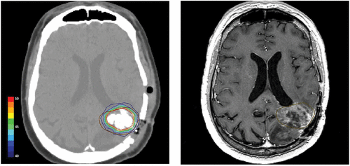
Figure 2. Postoperative CT scan of a glioblastoma patient showing magnetic nanoparticles deposits as hyperdense areas within the tumor tissue (left hand). Isothermal lines indicate calculated intratumoral temperatures for a magnetic field strength of 8.0 kA/m corresponding to an SAR of 718 W/kg (derived from the Hounsfield units in the CT scan). The brown line (right hand) represents the tumor area identified by preoperative MRI as fused with the CT scan.