Figures & data
Figure 1. Ultrasound of inflated balloon in mammary gland.
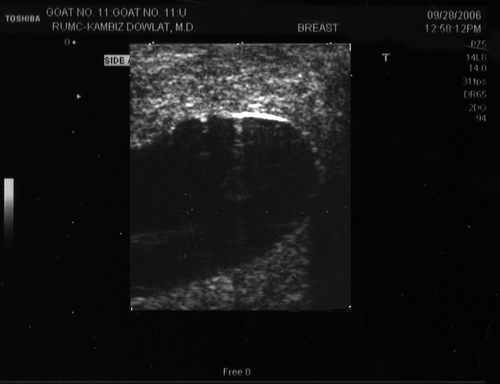
Figure 2. Intra-operative appearance of the inserted balloon and the subcutaneous thermistor.
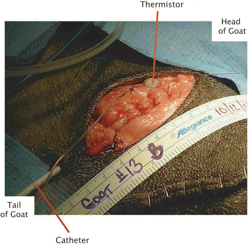
Figure 3. Sketch of a hot balloon as it lies inside the mammary gland.
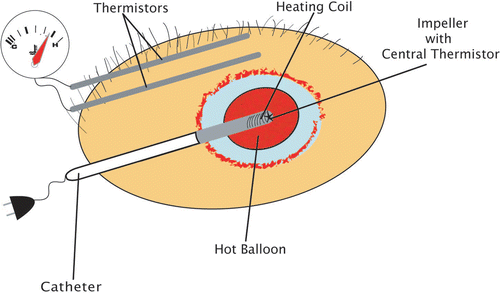
Figure 4. Balloon-catheter with heating coil at its center.
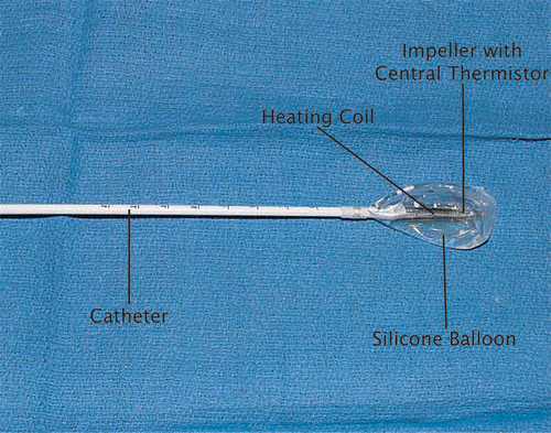
Figure 5. (A) Sketch of mammary gland sectioning at 3–4 mm intervals. (B) Representative section with radial sectioning at 3, 6, 9, and 12 o’clock.
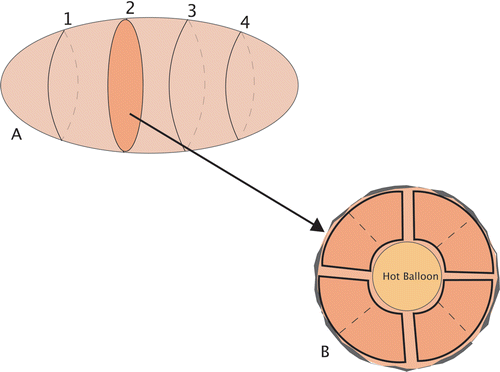
Figure 6. Histologic section of treated mammary gland. (A) Completely adjacent glandular structure immediately adjacent to the hot balloon. Underlying necrotic glands with normal appearing nuclei. (B) Glandular tissue with prominent new vessel formation, polymorphonuclear cells, and karyorrhectic debris. (C) Prominent new vessel formation in a background of myxoid stroma with mostly mononuclear cell infiltrate. No residual ductal glands are seen. (D) Morphologically normal mammary gland.
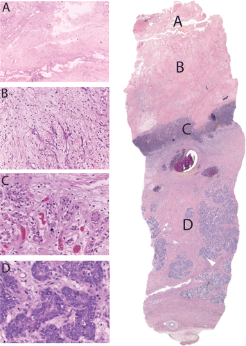
Figure 7. Average thermistor (sensor) readings (°C) for five glands treated for 15 minutes at 90°C balloon/central temperature.
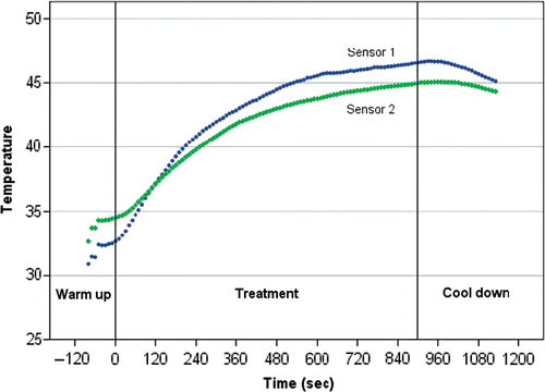
Figure 8. Gross appearance of two thermally treated mammary glands one week after thermotherapy.
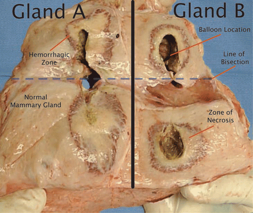
Figure 9. Mammary gland with superimposed histologic section.
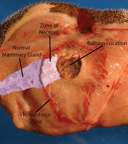
Table I. Data of treated goat mammary glands with treatment time and resulting mean necrosis during series I (FDA-approved ThermaChoice device).
Table II. Data of treated goat mammary glands with treatment time and resulting mean necrosis during series II (modified ThermaChoice device).
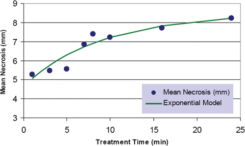
Figure 10. Modeling necrosis depth as a function of time.