907
Views
32
CrossRef citations to date
0
Altmetric
Original

Preliminary experience using high intensity focused ultrasound for treating liver metastasis from colon and stomach cancer

, , MD, , , , & show all
Pages 180-188 | Received 21 Dec 2007, Accepted 22 Nov 2008, Published online: 09 Jul 2009

Figures & data

Table I.  Summary of findings in colon cancer group (10 cases). All patients had chemotherapy treatment.

Figure 1. Picture of the HIFU device during an ablation procedure. The patient is under general anesthesia and in the prone position for treatment of metastatic tumor in the left lobe of the liver.
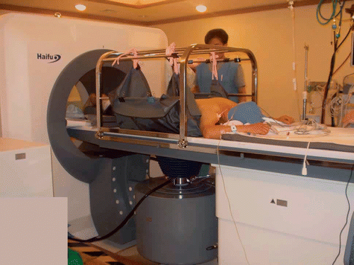
Figure 1. Picture of the HIFU device during an ablation procedure. The patient is under general anesthesia and in the prone position for treatment of metastatic tumor in the left lobe of the liver.

Table II.  Summary of findings in stomach cancer group. All patients had chemotherapy treatment.

Figure 2. Targets were ablated using mostly multiple HIFU row ablation zones with single ellipsoidal HIFU ablation zones when necessary. Deep portions were targeted first with shallower areas targeted subsequently.
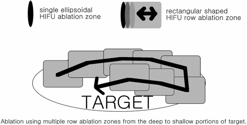
Figure 2. Targets were ablated using mostly multiple HIFU row ablation zones with single ellipsoidal HIFU ablation zones when necessary. Deep portions were targeted first with shallower areas targeted subsequently.

Table III.  Classification of results into four categories.

Figure 3. A 63-year-old man with multiple hepatic metastasis from colon carcinoma. Initial contrast enhanced coronal T1-weighted MR image shows one (arrow) of three metastatic nodules to the right lobe of liver (a). Ultrasonographic images showing the target before (b, left image) and after HIFU ablation (b, right image) demonstrate the hyperechoic changes representing optimal ablation. Follow-up contrast-enhanced coronal T1-weighted image two weeks after HIFU shows complete coagulative necrosis of the lesion (arrow). Pericostal ablation is noted in the sonification port (arrowheads) (c). Further follow-up contrast-enhanced CT 114 days after HIFU shows interval increase of the tumor size (arrows), indicating recurrence (d).
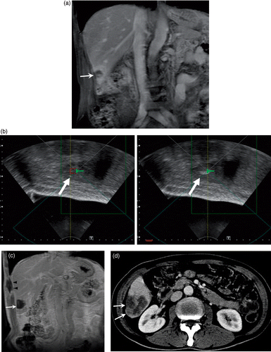
Figure 3. A 63-year-old man with multiple hepatic metastasis from colon carcinoma. Initial contrast enhanced coronal T1-weighted MR image shows one (arrow) of three metastatic nodules to the right lobe of liver (a). Ultrasonographic images showing the target before (b, left image) and after HIFU ablation (b, right image) demonstrate the hyperechoic changes representing optimal ablation. Follow-up contrast-enhanced coronal T1-weighted image two weeks after HIFU shows complete coagulative necrosis of the lesion (arrow). Pericostal ablation is noted in the sonification port (arrowheads) (c). Further follow-up contrast-enhanced CT 114 days after HIFU shows interval increase of the tumor size (arrows), indicating recurrence (d).

Figure 4. A 50-year-old woman with multiple hepatic metastasis from stomach carcinoma. Initial contrast-enhanced axial T1-weighted MR image shows one of multiple metastatic nodules in both lobes of the liver (a). Follow-up T1-weighted image two weeks after HIFU shows complete coagulative necrosis of the lesions (b). Although further follow up 107 days after HIFU shows progression of new lesions at non-ablated portions of the liver, no recurrence was seen at the ablated sites and decrease in size of the ablated sites are seen (c).
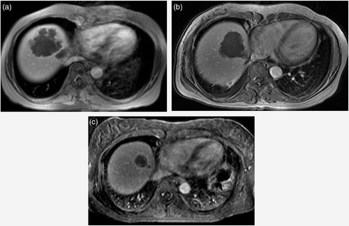
Figure 4. A 50-year-old woman with multiple hepatic metastasis from stomach carcinoma. Initial contrast-enhanced axial T1-weighted MR image shows one of multiple metastatic nodules in both lobes of the liver (a). Follow-up T1-weighted image two weeks after HIFU shows complete coagulative necrosis of the lesions (b). Although further follow up 107 days after HIFU shows progression of new lesions at non-ablated portions of the liver, no recurrence was seen at the ablated sites and decrease in size of the ablated sites are seen (c).

Reprints and Corporate Permissions

Please note: Selecting permissions does not provide access to the full text of the article, please see our help page How do I view content?

To request a reprint or corporate permissions for this article, please click on the relevant link below:

Academic Permissions

Please note: Selecting permissions does not provide access to the full text of the article, please see our help page How do I view content?

Obtain permissions instantly via Rightslink by clicking on the button below:

If you are unable to obtain permissions via Rightslink, please complete and submit this Permissions form. For more information, please visit our Permissions help page.