Figures & data
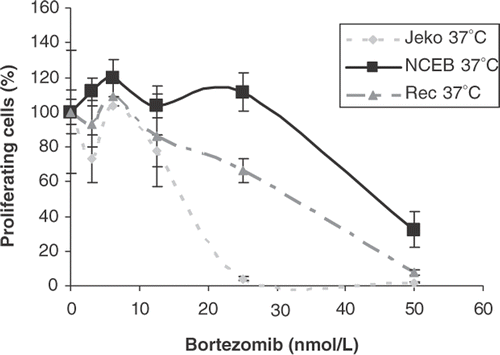
Figure 1. Cell survival expressed as percentage of proliferating cells of Jeko-1, Rec-1 and NCEB-1 in the presence of increasing concentrations of bortezomib. Cells were incubated with bortezomib (from 0 to 100 nmol/L) for 24 h in a humidified atmosphere at 37°C. Cells were harvested and the relative number of proliferating cells after treatment was assessed by standard WST-1 assay. Error bars represent mean ± standard deviation of triplicates in one representative experiment.

Table I. Kinetics of viability in MCL and haematological control cell lines following bortezomib treatment (left column). Cells were incubated with 25 nM of bortezomib, harvested at 24, 48 and 72 h and assessed for viability. The right column lists the IC50 values derived from the proliferating activity assessed by WST-1 assay in all cell lines treated for 24 h with increasing concentrations of bortezomib.
Table II. Survival rate in Jeko-1, Rec-1 and NCEB-1 after heat treatment at a mild thermal dose (41.8°C/120 min, left column) and at a severe thermal dose (44°C/30 min, right column). Cells were first heated and allowed to recover at 37°C in a humidified athmosphere for 24 h. Proliferation rate was evaluated by a standard WST assay.
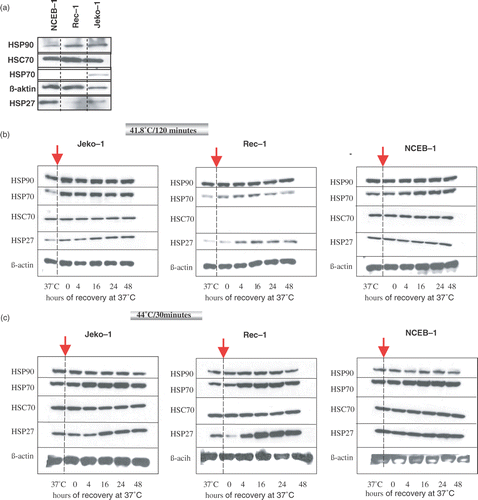
Figure 2. Expression level of hsp27, hsp70, Hsc70, and hsp90 by western blot analysis in an equal number of Jeko-1, Rec-1, and NCEB-1 cells at the physiological growth temperature of 37°C (a). Kinetics of expression of hsp27, hsp70, Hsc70, and hsp90 in Jeko-1, Rec-1 and NCEB-1 after exposure to a mild thermal dose (41.8°C/120 min) (b), and to 44°C/30 min (c). ß-actin was used as a protein loading control. After heat treatment at the selected thermal doses cells were allowed to recover in a humidified atmosphere at 37°C for up to 48 h. At the time points 4, 16, 24 and 48 h an equal numbers of cells were harvested for western blot analysis. Control cells were kept at 37°C (left column of each panel).

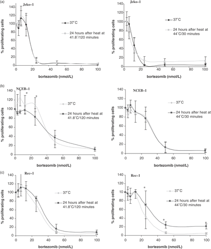
Figure 3. Cell survival expressed as percentage of proliferating cells in Jeko-1 (a), NCEB-1 (b), and Rec-1 (c), treated with increasing concentrations of bortezomib after a pre-treatment with heat at 41.8°C/120 min or 44°C/30 min. Briefly, cells were first heated, then allowed to recover for 24 h in a humidified atmosphere at 37°C. At this point cells were treated with bortezomib (from 0 to 100 nmol/L) for 24 h and then harvested for survival assessment by standard WST-1. Survival is expressed as a percentage of control cells (bortezomib-untreated) and compared with non-heated cells (37°C). Error bars represent mean ± standard deviation of three independent experiments. The percentage of proliferating cells at each concentration of bortezomib tested did not significantly differ between heated and non-heated cells and p-value was largely >0.5 (*).

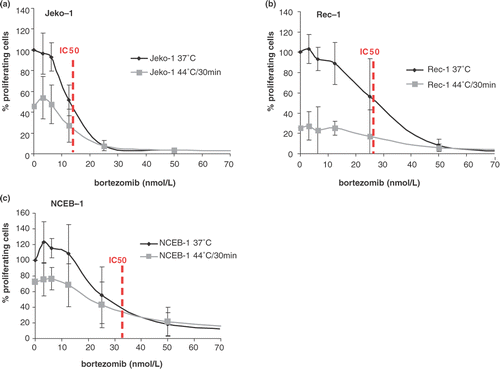
Figure 4. Cell survival expressed as a percentage of proliferating cells in Jeko-1 (a), Rec-1 (b), and NCEB-1 (c), treated with increasing concentrations of bortezomib given at 37°C (black line) or given concurrently to heat at 44°C/30 min (grey line). Survival of heated cells is expressed as a percentage of untreated cells at 37°C. Briefly, cells were first treated with bortezomib (from 0 to 100 nmol/L) and immediately heated. After the concomitant treatment, cells were incubated for 24 h in a humidified atmosphere at 37°C. At this point cells were harvested for survival assessment by standard WST-1. Error bars represent mean ± standard deviation of four independent experiments. Dotted lines indicated the IC50.

Table III. IC50 values obtained in Jeko-1, Rec-1, and NCEB-1 cells in the presence of increasing concentrations of bortezomib (from 0 to 100 nmol/L) for 24 h at: 37°C (control cells) listed in the left column; concomitant to heat treatment (middle column); or 24 h after pre-treatment with heat at the indicated thermal doses (right column).