Figures & data
Figure 1. This figure is an example showing the results of one batch of four mice, one in each treatment group, where an increased viral dissemination was noted in the heated tumor as compared to the non-heated tumor. Pixel frequency distribution was obtained from digitized auto-radiographs of the sectioned tumors. Background was determined based on the ‘No virus, no heat’ curve. Values above this background were considered evidence of gene expression or increased uptake in the tumor section. The number of pixels demonstrating activity above the background were quantified and expressed as a percentage of the total tumor area. Similar plots were obtained for all the batches studied to compare spread of virus.
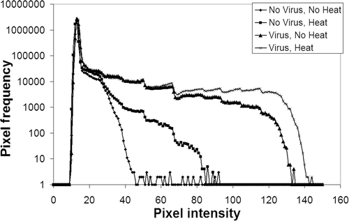
Figure 2. In vitro experiments were done to assess whether addition of heat at 41°C for 60 min would increase gene expression in transfected A549 cells. Various sequences of heating and viral infection were performed. There was no increased uptake of noted in any group as compared to the ‘Virus, no heat’ group. Group 1: No virus, no heat; group 2: No virus, heat; group 3: Virus, no heat; group 4: Virus → heat 1 h later; group 5: Heated virus, no cell heat; group 6: Virus → heat 24 h later; group 7: Heat → viral infection 1 h later; group 8: Heat → viral infection 6 h later.
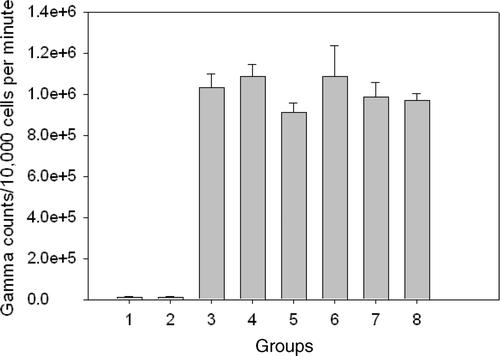
Table I. In vivo hyperthermia experiment where 1010 viral particles of AdhNIS were injected into the tumors followed 10 min later by HT at 41°C for 60 min. Area occupied by transgene expression in the tumor sections was compared. The numbers in the cells are the area occupied by the positive transgene expression as a percentage of the total area of the tumor section. In batch 4 increased area of expression was seen in the heated group and in batches 1, 2 and 3 increased area was noted in the non-heated tumors.
Table II. In vivo hyperthermia experiment where 1010 viral particles of AdhNIS were injected into the tumors 15 min following HT at 41°C for 60 min. Area occupied by transgene expression in the tumor sections was compared. The numbers in the cells are the area occupied by the positive transgene expression as a percentage of the total area of the tumor section. In batches 1, 3, 4 and 5 increased area of expression was seen in the heated group and in batches 2 and 6 increased area was noted in the non-heated tumors.
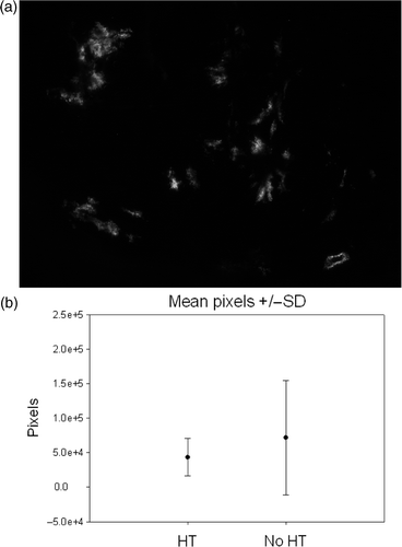
Figure 3. In vivo experiments were performed using an eGFP containing adenoviral vector. Intratumoral injection of the vector was performed 6 h after HT at 42°C for 30 min. (a) Example of green fluorescence as seen in one of the tumor sections. Total number of pixels in approximately 25–30 such images, 100 µm thick after every 200 µm, was quantified for each tumor (173 images for HT group and 162 for no HT group). (b) Box-plots of mean ± standard deviation showed no difference in the amount of fluorescence in the two groups (p = 0.17).