308
Views
9
CrossRef citations to date
0
Altmetric
Original

Conductive interstitial thermal therapy (CITT) inhibits recurrence and metastasis in rabbit VX2 carcinoma model

, DSc, , , , , , & show all
Pages 446-454 | Received 09 Feb 2009, Accepted 02 May 2009, Published online: 09 Sep 2009

Figures & data

Figure 1. Image of the VX2 carcinoma on a donor rabbit.

Figure 1. Image of the VX2 carcinoma on a donor rabbit.

Figure 2. The cavity created for accommodating the CITT probe prior to placement of the device (A), after placing the CITT device (B) and at the end of the CITT ablation (C).

Figure 2. The cavity created for accommodating the CITT probe prior to placement of the device (A), after placing the CITT device (B) and at the end of the CITT ablation (C).

Figure 3. A carcinoma mass developed after partial resection of the primary tumour in control group.
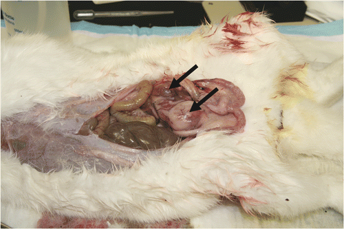
Figure 3. A carcinoma mass developed after partial resection of the primary tumour in control group.

Figure 4. The ablation site at 10 weeks post-CITT treatment showing no signs of scarring. The skin (A) and muscle (B) healed.

Figure 4. The ablation site at 10 weeks post-CITT treatment showing no signs of scarring. The skin (A) and muscle (B) healed.

Figure 5. H&E staining of histological slides that were taken from tissue collected at the ablation site at 10 weeks post CITT treatment. Complete dermal healing (A). Dense mature fibrous tissue (arrow) is present in healed dermis. Adjacent dermis (star) is normal. Residual inflammation was present in 10/12 animals (B, C, D). (B) Cellular debris and inflammatory cells (star) are walled off by a capsule of fibrous connective tissue (arrow). (C) Fragments of skeletal myofibres (arrow) are surrounded by necrotic cellular debris and inflammatory cells. (D) Inflammatory infiltrate surrounding myofibre. H&E, magnification 20× (A), 40× (B), 200× (C) and 400× (D).
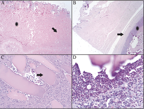
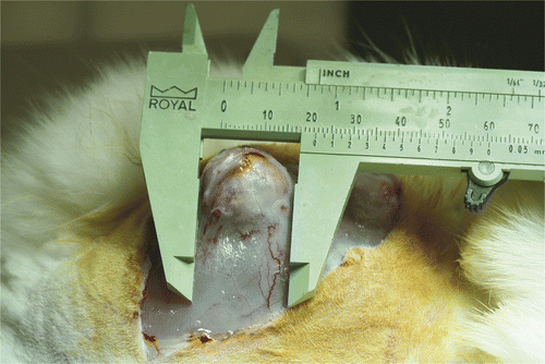
Figure 5. H&E staining of histological slides that were taken from tissue collected at the ablation site at 10 weeks post CITT treatment. Complete dermal healing (A). Dense mature fibrous tissue (arrow) is present in healed dermis. Adjacent dermis (star) is normal. Residual inflammation was present in 10/12 animals (B, C, D). (B) Cellular debris and inflammatory cells (star) are walled off by a capsule of fibrous connective tissue (arrow). (C) Fragments of skeletal myofibres (arrow) are surrounded by necrotic cellular debris and inflammatory cells. (D) Inflammatory infiltrate surrounding myofibre. H&E, magnification 20× (A), 40× (B), 200× (C) and 400× (D).

Reprints and Corporate Permissions

Please note: Selecting permissions does not provide access to the full text of the article, please see our help page How do I view content?

To request a reprint or corporate permissions for this article, please click on the relevant link below:

Academic Permissions

Please note: Selecting permissions does not provide access to the full text of the article, please see our help page How do I view content?

Obtain permissions instantly via Rightslink by clicking on the button below:

If you are unable to obtain permissions via Rightslink, please complete and submit this Permissions form. For more information, please visit our Permissions help page.