894
Views
23
CrossRef citations to date
0
Altmetric
Original

Heat shock increases expression of NAD(P)H:quinone oxidoreductase (NQO1), mediator of β-lapachone cytotoxicity, by increasing NQO1 gene activity and via Hsp70-mediated stabilisation of NQO1 protein

, , , , , , & , MD, PhD show all
Pages 477-487 | Received 16 Feb 2009, Accepted 18 May 2009, Published online: 09 Sep 2009

Figures & data

Figure 1. NQO1 expression and cell death caused by β-lap. (A) Effects of β-lap on clonogenic survival of MDA-MB-231(NQO1+) and MDA-MB-231(NQO1−) cells. As shown in the inset, NQO1 is over-expressed in MDA-MB-231(NQO1+) cells, while NQO1 expression in MDA-MB-231(NQO1−) cells is negligible. Cells were treated with different concentrations of β-lap for 4 h at 37°C, washed, cultured for 10–14 days, and the colonies were counted. An average of seven experiments ± SE is shown. (B) Effects of preheating on the sensitivity of MDA-MB-231(NQO1+) and MDA-MB-231(NQO1−) cells to β-lap. Cells were heated at 42°C for 1 h, incubated at 37°C for varying lengths of time, and treated with 5 µM β-lap for 4 h. After β-lap treatment, cells were washed, cultured for 10–14 days, and the colonies were counted. An average of six experiments ± SE is shown.
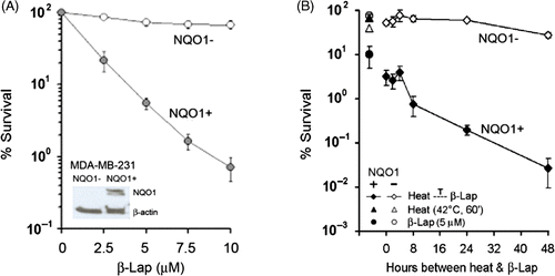
Figure 1. NQO1 expression and cell death caused by β-lap. (A) Effects of β-lap on clonogenic survival of MDA-MB-231(NQO1+) and MDA-MB-231(NQO1−) cells. As shown in the inset, NQO1 is over-expressed in MDA-MB-231(NQO1+) cells, while NQO1 expression in MDA-MB-231(NQO1−) cells is negligible. Cells were treated with different concentrations of β-lap for 4 h at 37°C, washed, cultured for 10–14 days, and the colonies were counted. An average of seven experiments ± SE is shown. (B) Effects of preheating on the sensitivity of MDA-MB-231(NQO1+) and MDA-MB-231(NQO1−) cells to β-lap. Cells were heated at 42°C for 1 h, incubated at 37°C for varying lengths of time, and treated with 5 µM β-lap for 4 h. After β-lap treatment, cells were washed, cultured for 10–14 days, and the colonies were counted. An average of six experiments ± SE is shown.

Figure 2. Expression of NQO1 mRNA and NQO1 protein production upon heat treatment. A549 and MDA-MB 231 cells (NQO1+) were heated at 42°C for 1 h, incubated at 37°C, and harvested at the times indicated. (A) The expression of NQO1 mRNA was analysed by RT-PCR. (B) Protein expression was determined by Western blot. Hsp70 is shown as a relevant marker for heat treatment. Each experiment was independently performed three times.
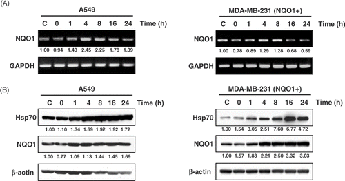
Figure 2. Expression of NQO1 mRNA and NQO1 protein production upon heat treatment. A549 and MDA-MB 231 cells (NQO1+) were heated at 42°C for 1 h, incubated at 37°C, and harvested at the times indicated. (A) The expression of NQO1 mRNA was analysed by RT-PCR. (B) Protein expression was determined by Western blot. Hsp70 is shown as a relevant marker for heat treatment. Each experiment was independently performed three times.

Figure 3. Changes of NQO1 promoter activities in A549 and MDA-MB 231 cells by heat treatment. (A) NQO1 promoter-luciferase constructs were generated for transfection. Upstream promoter regions were generated by PCR. (B) Relative luciferase activities from various NQO1 promoter deletions in A549 and MDA-MB-231 cells. Five micrograms of test plasmid (pGL3-NQOp) and 1 µg of pRL-SV40 were co-transfected into A549 and MDA-MB 231 (NQO1+) cells. After 18 h incubation, the transfected cells were heated at 42°C for 1 h, and harvested for dual luciferase reporter assay. Data are expressed as relative luciferase activity to those of the construct, pGL3-NQO1p5. Promoter activity values represent the mean of three independent experiments.
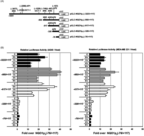
Figure 3. Changes of NQO1 promoter activities in A549 and MDA-MB 231 cells by heat treatment. (A) NQO1 promoter-luciferase constructs were generated for transfection. Upstream promoter regions were generated by PCR. (B) Relative luciferase activities from various NQO1 promoter deletions in A549 and MDA-MB-231 cells. Five micrograms of test plasmid (pGL3-NQOp) and 1 µg of pRL-SV40 were co-transfected into A549 and MDA-MB 231 (NQO1+) cells. After 18 h incubation, the transfected cells were heated at 42°C for 1 h, and harvested for dual luciferase reporter assay. Data are expressed as relative luciferase activity to those of the construct, pGL3-NQO1p5. Promoter activity values represent the mean of three independent experiments.

Figure 4. Effects of Act D and CHX on NQO1 mRNA and protein expression, respectively, in A549 and MDA-MB 231 (NQO1+) cells. Cells were heated at 42°C for 1 h, incubated at 37°C for 4 h, and treated with 4 μg/mL Act D or 50 μg/mL CHX for the indicated times. (A) NQO1 mRNA expression was determined by RT-PCR. (B) NQO1 protein expression was determined by Western blot.
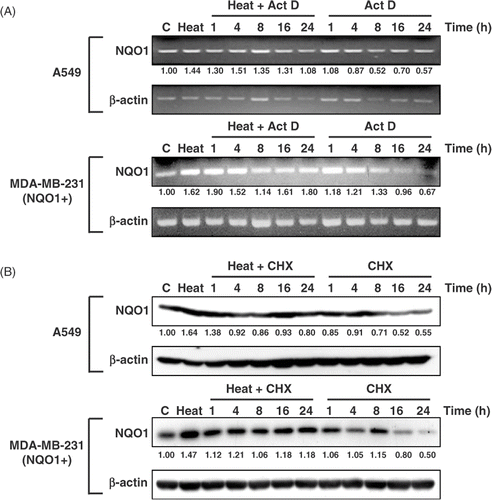
Figure 4. Effects of Act D and CHX on NQO1 mRNA and protein expression, respectively, in A549 and MDA-MB 231 (NQO1+) cells. Cells were heated at 42°C for 1 h, incubated at 37°C for 4 h, and treated with 4 μg/mL Act D or 50 μg/mL CHX for the indicated times. (A) NQO1 mRNA expression was determined by RT-PCR. (B) NQO1 protein expression was determined by Western blot.

Figure 5. Co-immunoprecipitation and localisation of NQO1 and Hsp70. (A) A549 and MDA-MB 231 cells (NQO1+) were harvested at 8 h after heating at 42°C for 1 h. Whole cell lysates were immunoprecipitated with mouse anti-Hsp70 antibody or mouse anti-NQO1 antibody. The precipitates were analysed by SDS-PAGE and blotted onto polyvinylidene fluoride membranes. The membranes were probed with rabbit anti-NQO1 antibody, or rabbit anti-Hsp70 antibody and binding was revealed by enhanced chemiluminescence. (B) A549 cell lysate was precipitated with anti-Hsp90 antibody or anti-Hsp40 antibody as previously described. (C) MDA-MB-231 (NQO1+) cells were transfected with siHsp70 or control siRNA and incubated 48h. Then the cells were heated at 42°C for 1 h and incubated for various times at 37°C. Expression of NQO1 and Hsp70 protein were analysed by Western blot. (D) Cells were heated at 42°C for 1 h, incubated for 8 h at 37°C, and then labelled for NQO1 (red) and Hsp70 (green) using FITC-conjugated anti-rabbit secondary antibody and Texas-Red conjugated anti-mouse secondary antibody, respectively. Labelled cells were examined using confocal microscopy.
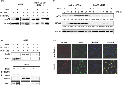
Figure 5. Co-immunoprecipitation and localisation of NQO1 and Hsp70. (A) A549 and MDA-MB 231 cells (NQO1+) were harvested at 8 h after heating at 42°C for 1 h. Whole cell lysates were immunoprecipitated with mouse anti-Hsp70 antibody or mouse anti-NQO1 antibody. The precipitates were analysed by SDS-PAGE and blotted onto polyvinylidene fluoride membranes. The membranes were probed with rabbit anti-NQO1 antibody, or rabbit anti-Hsp70 antibody and binding was revealed by enhanced chemiluminescence. (B) A549 cell lysate was precipitated with anti-Hsp90 antibody or anti-Hsp40 antibody as previously described. (C) MDA-MB-231 (NQO1+) cells were transfected with siHsp70 or control siRNA and incubated 48h. Then the cells were heated at 42°C for 1 h and incubated for various times at 37°C. Expression of NQO1 and Hsp70 protein were analysed by Western blot. (D) Cells were heated at 42°C for 1 h, incubated for 8 h at 37°C, and then labelled for NQO1 (red) and Hsp70 (green) using FITC-conjugated anti-rabbit secondary antibody and Texas-Red conjugated anti-mouse secondary antibody, respectively. Labelled cells were examined using confocal microscopy.

Reprints and Corporate Permissions

Please note: Selecting permissions does not provide access to the full text of the article, please see our help page How do I view content?

To request a reprint or corporate permissions for this article, please click on the relevant link below:

Academic Permissions

Please note: Selecting permissions does not provide access to the full text of the article, please see our help page How do I view content?

Obtain permissions instantly via Rightslink by clicking on the button below:

If you are unable to obtain permissions via Rightslink, please complete and submit this Permissions form. For more information, please visit our Permissions help page.