906
Views
60
CrossRef citations to date
0
Altmetric
Original Article

Ultrasound-guided percutaneous microwave ablation of adrenal metastasis: Preliminary results

, , MD, , , &
Pages 455-461 | Received 10 Feb 2009, Accepted 25 May 2009, Published online: 09 Sep 2009

Figures & data

Table 1.  Summary of patients and tumours treated with MW ablation.

Figure 1. Ultrasound-guided percutaneous MW ablation in a 53-year-old man with right adrenal metastasis (arrow) from hepatocellular carcinoma. (a) Greyscale ultrasound shows a hypoechoic tumour in the right adrenal gland before MW ablation. (b) The tumour (arrow) showed rich enhancement on contrast-enhanced ultrasound. (c) MW antenna (arrowhead) was percutaneously placed into the adrenal tumour (arrow) under real-time ultrasound guidance. (d) The tumour (arrow) became hyperechoic right after MW ablation.
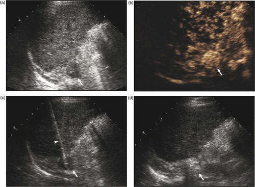
Figure 1. Ultrasound-guided percutaneous MW ablation in a 53-year-old man with right adrenal metastasis (arrow) from hepatocellular carcinoma. (a) Greyscale ultrasound shows a hypoechoic tumour in the right adrenal gland before MW ablation. (b) The tumour (arrow) showed rich enhancement on contrast-enhanced ultrasound. (c) MW antenna (arrowhead) was percutaneously placed into the adrenal tumour (arrow) under real-time ultrasound guidance. (d) The tumour (arrow) became hyperechoic right after MW ablation.

Figure 2. MW ablation in a 76-year-old woman who had a right adrenal metastasis from clear-cell renal carcinoma two years after right nephrectomy. (a) Before treatment the tumour was slightly enhanced on contrast-enhanced MRI. No enhancement was observed on contrast-enhanced MRI at three months (b), one year (c) and two years (d) after MW ablation, the ablated tumour gradually shrank over time.
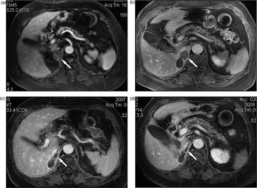
Figure 2. MW ablation in a 76-year-old woman who had a right adrenal metastasis from clear-cell renal carcinoma two years after right nephrectomy. (a) Before treatment the tumour was slightly enhanced on contrast-enhanced MRI. No enhancement was observed on contrast-enhanced MRI at three months (b), one year (c) and two years (d) after MW ablation, the ablated tumour gradually shrank over time.

Figure 3. MW ablation in a 51-year-old man who had a left adrenal metastasis from hepatocellular carcinoma. (a) Before treatment the tumour was enhanced on contrast-enhanced MRI. No enhancement was observed on contrast-enhanced MRI at three months (b), six months (c) and one year (d) after MW ablation.
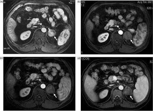
Figure 3. MW ablation in a 51-year-old man who had a left adrenal metastasis from hepatocellular carcinoma. (a) Before treatment the tumour was enhanced on contrast-enhanced MRI. No enhancement was observed on contrast-enhanced MRI at three months (b), six months (c) and one year (d) after MW ablation.

Reprints and Corporate Permissions

Please note: Selecting permissions does not provide access to the full text of the article, please see our help page How do I view content?

To request a reprint or corporate permissions for this article, please click on the relevant link below:

Academic Permissions

Please note: Selecting permissions does not provide access to the full text of the article, please see our help page How do I view content?

Obtain permissions instantly via Rightslink by clicking on the button below:

If you are unable to obtain permissions via Rightslink, please complete and submit this Permissions form. For more information, please visit our Permissions help page.