Figures & data
Table 1. Distribution of primary tumours.
Figure 1. Transverse images of a 75-year-old woman with liver metastasis of colorectal cancer origin treated with low-frequency microwave ablation (LF-MWA). (a) MR image (T1 weighted) obtained 2 weeks before MWA shows a single metastasis (arrow) in segment 4 with a maximum diameter of 31.5 mm and a volume of 10.39 mL. (b) Computed tomography (CT) image obtained during MWA (30 min, 45 W). (c) MR image (T1 weighted) obtained 24 h after successful treatment shows typical coagulation (volume: 20.23 mL, max. diameter: 44.3 mm). (d) MR image obtained 48 months (volume: 5.11 mL) after MWA shows area of scarring in the ablation zone with no progress in size.
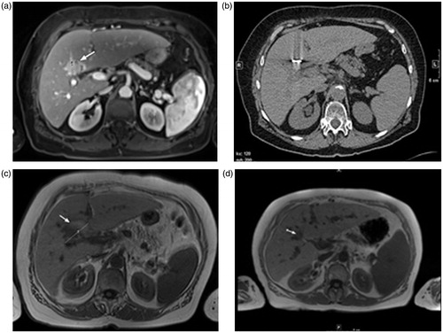
Figure 2. Transverse images of a 40-year-old man with liver metastasis from colorectal cancer treated with high-frequency microwave ablation (HF-MWA). (a) MR image (T2 weighted) obtained 4 weeks prior to MWA shows metastasis (arrow) in segment 5 with a maximum diameter of 17.3 mm and a volume of 9.07 mL. (b) Computed tomography (CT) image obtained during MWA (5 min, 76 W). (c) MR image (T1 weighted) obtained 24 h after successful treatment shows typical coagulation (volume: 23.19 mL, max. diameter: 9.02 mm). (d) MR image (T1 weighted) 48 months (volume: 2.18 mL) after MWA shows area of scarring and no progression of the tumour.
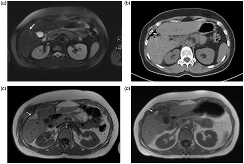
Table 2. Coagulation volume in millilitres and volume decrease after ablation in percentages in parentheses.
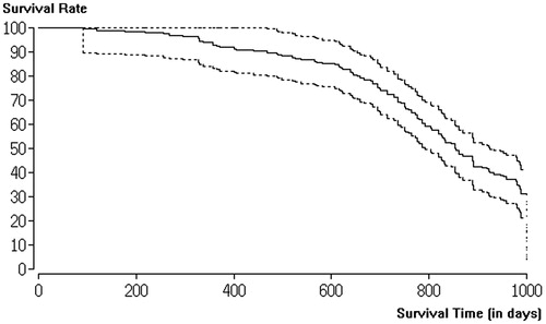
Figure 3. Kaplan–Meier curve of patients treated with low-frequency microwave ablation (LF-MWA) for palliative indication.
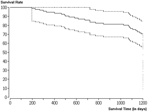
Figure 4. Kaplan–Meier curve of patients treated with high-frequency microwave ablation (HF-MWA) for palliative indication.