
Figures & data
Figure 1. High intensity focused ultrasound (HIFU) treatment of breast fibroadenoma using the Echopulse device (Theraclion Ltd., Malakoff, France).
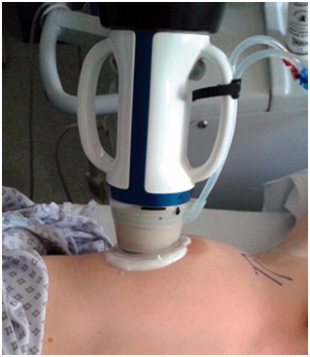
Figure 2. Treatment planning and final treatment. From left to right: (1) manual drawing of outline of fibroadenoma (FAD) (blue) and skin (red) on touchscreen unit in anti-radial position and number of treatment pulses (white) calculated by the Echopulse; (2) radial view of target volume with treatment pulses (white cylinders) calculated by the Echopulse; (3) application of treatment pulse in centre of FAD; (4) final treatment of two circumferential rings, showing completed pulses (green) and deselected pulses (grey).
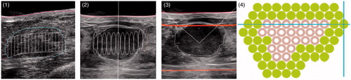
Figure 3. Box and whisker plot for high intensity focused ultrasound (HIFU) treatment and control group (untreated) demonstrating not significant difference in pre-treatment volume (blue) and age (green) of the patients.
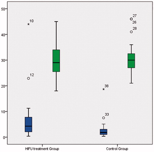
Figure 4. Histopathology of excised fibroadenoma treated with high intensity focused ultrasound (HIFU) showing fibrous scarring on low (left) and high power (right).
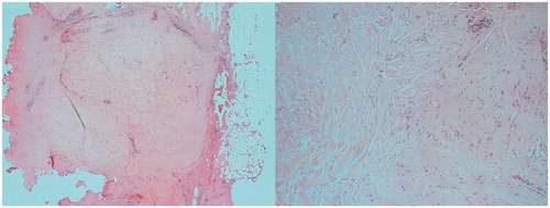
Figure 5. Short-term complications (a, b) two images of ecchymosis at 2 weeks, (c) hyper-pigmentation at 3 months and (d) first-degree skin burn at 2 weeks post-treatment.
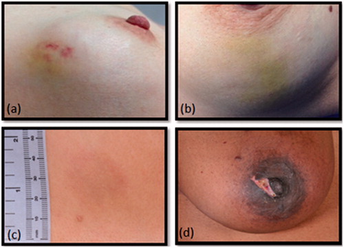
Figure 6. Change in volume per patient at 2-week, 3- and 6-month follow-up (in %).
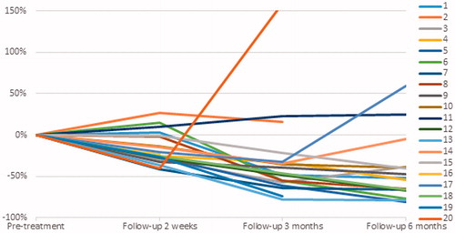
Table 1. Volume changes during 6-month follow-up in (a) high intensity focused ultrasound (HIFU) treatment group and (b) control group.