Figures & data
Table 1. The clinical characteristics and outcomes of RFA in the 6 patients with low risk small papillary thyroid carcinoma.
Figure 1. Volume changes of ablation zones after radiofrequency ablation of papillary thyroid carcinomas.
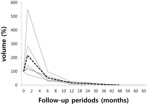
Figure 2. (A, B and C) Complete disappearance of papillary thyroid carcinoma after radiofrequency ablation. (A) Ultrasonography of a 64-year-old woman revealed a 10 mm mass proven as a papillary thyroid carcinoma on both fine-needle aspiration and core-needle biopsy in the right thyroid gland. (B) At 1-month follow-up after radiofrequency ablation, ultrasonography revealed a 12 mm ablation zone. (C) At 2-year follow-up, the ablation zone completely disappeared on ultrasonography.
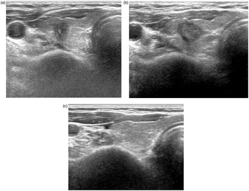
Figure 3. (A–E) Gradual reduction and complete disappearance of papillary thyroid carcinoma after radiofrequency ablation. (A) Ultrasonography of a 76-year-old woman revealed a 6-mm mass in the left thyroid gland. (B) FNA cytology of the mass revealed papillary thyroid carcinoma cells with nuclear atypia, including nuclear grooves, high N/C ratio and nuclear pseudoinclusion (Papanicolaou stain, original magnification ×400). (C) At 2-year follow-up after radiofrequency ablation, US revealed a smaller but persistent, 4-mm ill-defined ablation zone in the left thyroid gland. (D) FNA cytology of the ablation zone demonstrated benign follicular epithelial cells with hemosiderin pigment-laden macrophages (× 100). (E) At 4-year follow-up, US revealed complete disappearance of the ablation zone.
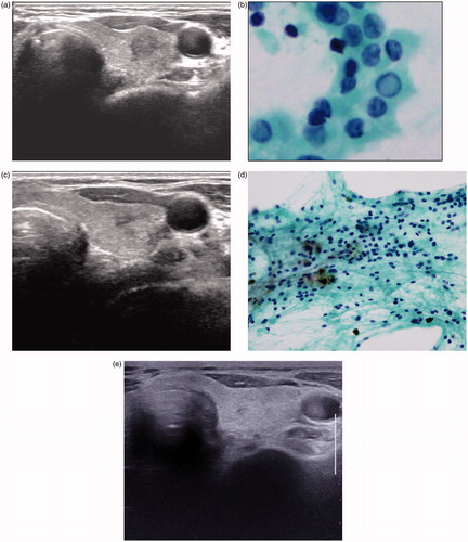
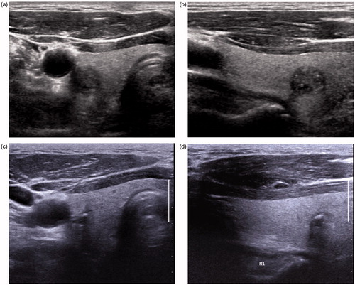
Figure 4. (A–D) Gradual reduction and resultant calcified residues of papillary thyroid carcinoma after radiofrequency ablation. (A and B) Transverse and longitudinal ultrasonography of a 79-year-old man revealed an 11-mm mass proven as a papillary thyroid carcinoma on-fine needle aspiration in the right thyroid gland. (C and D) At 4-year follow-up after radiofrequency ablation, transverse and longitudinal ultrasonography revealed a 3-mm calcified residue in the ablation zone. At 3 year, fine-needle aspiration and core-needle biopsy of the ablation zone revealed no tumour cells (results not shown).