
Figures & data
Figure 1. Flowchart of patient selection.
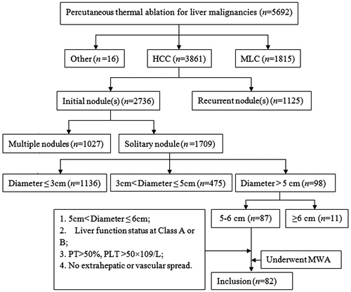
Table 1. Clinical characteristics of the 82 enrolled patients.
Figure 2. Imaging document of the presence and disappearance of HCC lesions. The MWA-treated subject was a 52-year-old male HCC patient who had HBV-related liver cirrhosis. (A) Arterial phase; (B) Portalvenous phase. Pre-treatment CT images shows a tumour as a 5.4-cm hyperintense nodule on an intense arterial enhancement (A), with an enhancement recession (B). (C) Arterial and (D) portalvenous phase. MRI images obtained at four weeks after treatment show no enhancements inside or beside the ablation zone.
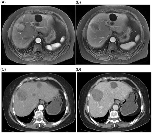
Table 2. Analysis of factors associated with local recurrence.
Figure 3. Recurrence-free survival curves after MWA treatment.
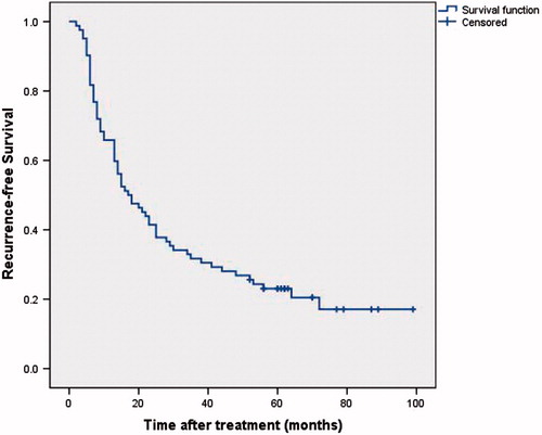
Figure 4. Overall survival curves after MWA treatment.
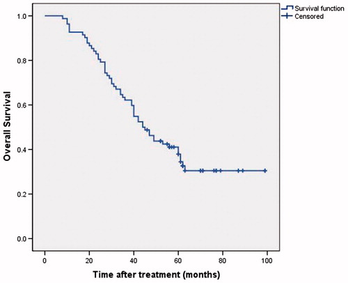
Table 3. Analysis of factors associated with overall survival.