
Figures & data
Figure 1. MWA treatment of a 35-year old man with a left thyroid solid nodule. (A) Two-dimensional ultrasonic image of the thyroid nodule. (B) The colour Doppler image of the thyroid nodule. (C) The contrast enhanced ultrasound image of the thyroid nodule before treatment. (D) The contrast enhanced ultrasound image of the nodule on day 3 after the ablation. (E) Two-dimensional ultrasonic image of the nodule three month after the ablation. (F) The colour Doppler image of the nodule three month after the ablation. Vascularity was shown around the nodule. (G) The colour Doppler image of the nodule six month after the ablation. Vascularity was shown inside the nodule in ablation area. (H) The contrast enhanced ultrasound image of the nodule six month after the ablation. Reperfusion was shown in the ablation area of the nodule.
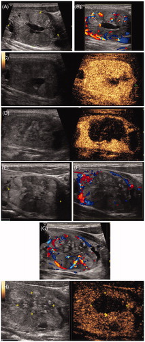
Figure 2. Hydrodissection technique. (A) On transverse image, the nodule is located adjacent to the common carotid artery. (B) 0.9 % normal saline is injected between the common carotid artery and the nodule to prevent needle-induced thermal injury. M = mass, CCA = common carotid artery. Arrowheads = microwave antenna.
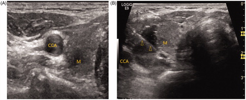
Table 1. The changes in volume before microwave ablation and at each follow-up.
Table 2. Comparison of effective therapy group versus recurrence group.