
Figures & data
Table 1. Demographics, size and location of the lesion.
Figure 1. Computed tomography axial scans of a patient with symptomatic intra-articular osteoid osteoma (biopsy proven). (A) White arrow illustrates the nidus (5 mm in diameter) of an intra-articular osteoid osteoma in the tibio-fibular joint. (B) Once inside the nidus, coaxially through the OnControl trocar the bone biopsy needle is inserted for sampling. (C) Post biopsy, the radiofrequency electrode is coaxially inserted and ablation session is performed with osteoid osteoma protocol according the manufacturer’s guidelines. (D) Post-ablation scan illustrating the access through normal bone ending inside the nidus of the osteoid osteoma.
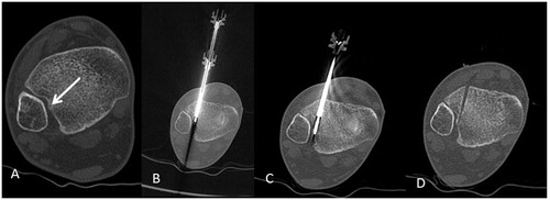
Figure 2. Computed tomography axial scans of a patient with symptomatic intra-articular osteoid osteoma in the hand (biopsy proven). (A) Radio-opaque mesh is placed over the skin for entry point selection. (B) Post biopsy, the radiofrequency electrode is coaxially inserted and ablation session is performed with osteoid osteoma protocol according the manufacturer’s guidelines.
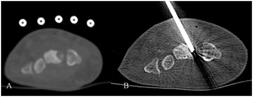
Table 2. Literature review of osteoid osteoma ablation techniques.