Figures & data
Figure 1. Ex-vivo gross examination of ventilated lungs after RFA. A: pleural surface detail showing the pleural hole of the needle tract. B: ex vivo examination with ventilated lungs showing the air leakage (bubbles).
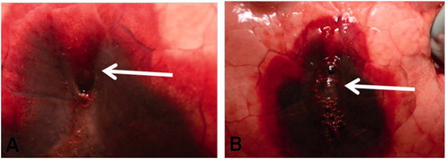
Figure 2. Scanning electron microscopy. Axial slice, showing a widely open tract (arrow). Condensation of the lung parenchyma around the tract (arrowheads).
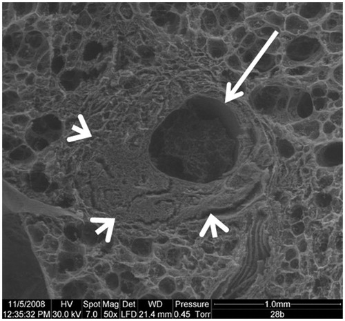
Figure 3. Optical Microscopy with orcein staining. Lung damages related to the needle positioning alone, without any thermo-ablation. A: Biopsy path without haemorrhagic modifications, peripheral parenchyma is preserved (mag ×50). B: Hematoxylin staining showing haemorrhagic modifications, with red blood cells in the alveoli (mag ×200).
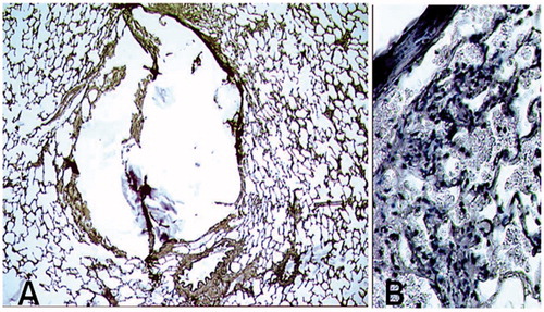
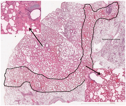
Figure 4. H&E staining. Concentric haemorrhagic modifications along the RFA needle. Left cartridge: centre with alveolar spaces filled by haemorrhagic fluid. Right cartridge: periphery with congestion of the capillaries.