Figures & data
Figure 1. In vivo experimental setup. Mice were placed on the positioning tray within the MR receive coil. Acoustic coupling was achieved by using distilled degassed water and degassed ultrasound gel. An acoustic absorber, coupled with ultrasound gel, was placed on the animal’s back to control the exit beam and minimise movement. Respiration was monitored by an MR-compatible small animal monitoring system.
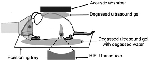
Figure 2. Planning and temperature mapping for image-guided hyperthermia. The KPC tumour was identified on the planning images in the tail of the pancreas (A). The ROI was placed in the centre of the tumour. A dashed line to outline the tumour has been added to show the reader the tumour border. Real-time MRI-based temperature monitoring using the PRFS method shown in colour overlaid on the planning image (B). Representative mean temperature in the target region during a sonication (C). Stable mild hyperthermia was achieved in the target region through binary feedback control of temperature. An algorithm was utilised to keep the temperature in the range of 41–42.5 °C (horizontal lines) within the ROI (4 mm diameter). Orange vertical lines represent the start and end of the sonication.
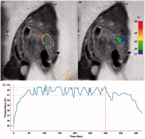
Figure 3. Box-and-Whisker plot of doxorubicin (Dox) concentration in the tumours of KPC mice treated with 15 mg Dox/kg low-temperature-sensitive liposomal doxorubicin (LTSL-Dox) and non-liposomal doxorubicin (Dox), with and without the application of MR-HIFU. *Denotes significance at the p < 0.05 level.
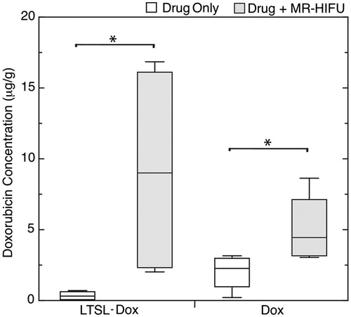
Table 1. Median doxorubicin concentration [interquartile range] measured in pancreatic tumours following different treatment regimens (n = 4 in each group).
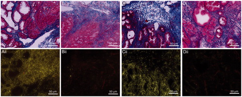
Figure 4. Representative images of serial sections stained with Masson’s trichrome (i) and fluorescent imaging (ii). Images demonstrate the distribution of doxorubicin and blood vessels within pancreatic tumours treated with LTSL-Dox + MR-HIFU (A); LTSL alone (B); DOX + MR-HIFU (C) OR Dox alone (D).