Figures & data
Figure 1. BRCA2 degradation in established cell lines of various tumour origins. Immunoblots of cells without treatment (37 °C) or treated with hyperthermia (42 °C) for 60 min effectively, excluding 15 min of pre-heating time. Upper row of the panels show the BRCA2-signal, the lower panel shows the PARP-1-signal. Each panel represents a set samples from a different cell line, from left to right: HeLa (cervical), FaDu (head- and neck), T24 (bladder) and MDA-MB-231 (breast). Percentages at the bottom of each panel indicate the relative intensity of the BRCA2 signal (corrected to PARP-1) in the heat-treated sample compared to the non-treated sample.
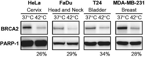
Figure 2. Schematic representation of tumour collection. Fresh biopsies obtained are dissociated enzymatically and mechanically. Dissociated suspensions of tumours collected were prepared for treatment by centrifugation (5 min, 300 rcf) and subsequent resuspension in RMPI supplemented with 10% FCS. The resulting cell suspension was divided over two separate culture dishes: one was incubated at 37 °C for 75 min in a controlled environment (37 °C, 5% CO2, 20% O2), while the other was incubated at 42 °C in a similarly controlled environment for the same amount of time, allowing 15 min to pre-heat the medium and thus resulting in 60 min of effective heating. After the treatment, cells in both samples are lysed and the proteins are analysed on an immunoblot.
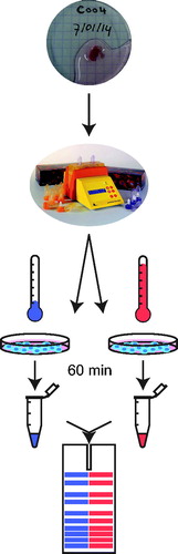
Figure 3. BRCA2 degradation in tumours heated ex vivo. Immunoblots of (A) 11 cervical tumours, (B) five head and neck tumours, and (C) 16 bladder tumours. Each pair of samples represents the non-hyperthermia-treated and treated samples of a tumour sample. ORC2 is shown as a loading control. Percentages at the bottom of each panel indicate the relative intensity of the BRCA2 signal (corrected to ORC2) in the heat-treated sample compared to the non-treated sample. *For clarity of presentation, the shadow values were reset from 0 to 175 in Adobe Photoshop. **PARP-1 is shown as a loading control and as an alternative to ORC2, because the latter could not be detected in this sample.
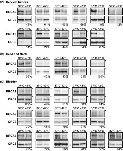
Figure 4. BRCA2 protein levels in biopsies taken before and after hyperthermia treatment in vivo. Immunoblots of biopsies taken before (pre) and after (post) hyperthermia in (A) breast tumours and in (B) cervical tumours. Samples marked with “42 °C” received ex vivo hyperthermia as an internal control. Shown are BRCA2 and ORC2. The percentages at the bottom of each panel indicate the relative intensity of the BRCA2 signal (corrected to ORC2) in the heat-treated sample compared to the non-treated sample. *For clarity of presentation the shadow values were reset from 0 to 175 in Adobe Photoshop.
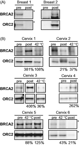
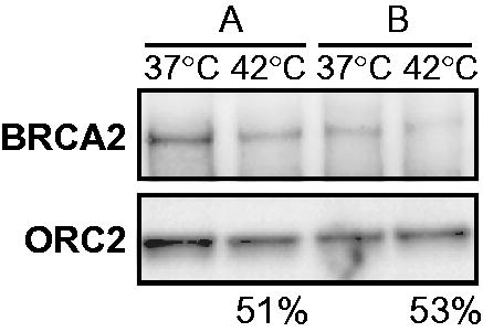
Figure 5. Tumour heterogeneity contributes to pre-hyperthermia protein levels of BRCA2 in cervical tumours. Immunoblots of two biopsies (A and B) taken from the same cervical tumour and treated ex vivo in the same hyperthermia-session at 42 °C. Upper panel shows the BRCA2-signal, the lower ORC2. The percentages at the bottom of the panel indicate the relative intensity of the BRCA2 signal (corrected to ORC2) in the heat-treated sample compared to the non-treated sample for each biopsy.