
Figures & data
Figure 1. Measurement of body composition parameters using axial CT slice at the level of the third lumbar vertebra. (a) Delineation of total muscle area (pink), using a threshold of −29 to +150 HU. (b) Delineation of visceral (green) and (c) subcutaneous (blue) fat areas, using a threshold of −190 to −30 HU.
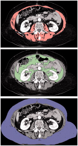
Table 1. Baseline demographic and disease characteristics of the 214 patients.
Figure 2. Overall survival curves regarding albumin rate.
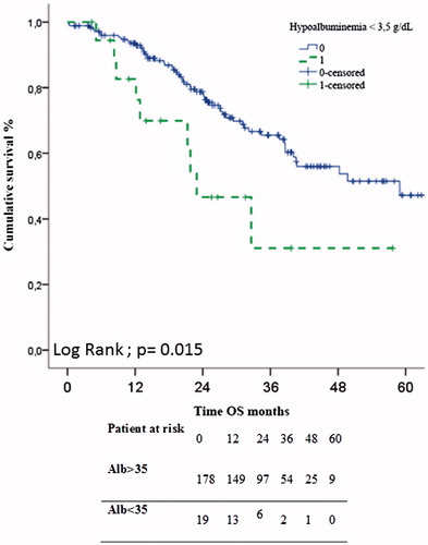
Table 2. Outcomes after cytoreductive surgery separated by nutritional factors in univariate analysis.
Table 3. Univariate and multivariate analyses of overall survival after cytoreductive surgery for colorectal peritoneal metastasis.
Table 4. Comparison of patients, with or without major complication, after cytoreductive surgery for peritoneal carcinomatosis separated by nutritional factor, n (%).