Figures & data
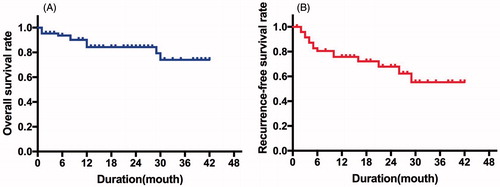
Figure 1. CEUS-guided ablation at needle tract. (A) CEUS showed continuous linear microbubble extravasated along the needle tract (white arrow). (B) Electrode was inserted into outer parts of needle tract which was near the capsule under the guidance of CEUS (white arrow). (C) CEUS showed no microbubble extravasation along the needle tract after thermal ablation. The high echo showed by white arrow was caused by the carbonisation of electrode, not needle tract bleeding.
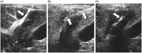
Table 1. The demographic and tumour information of patients included in this study.
Figure 2. Survival curves. (A) The OS rates were 84.8%, 82.7% and 82.7% at the 1-, 2-, and 3-year time points, respectively. (B) The RFS rates were 67.9%, 64.0% and 64.0% at the 1-, 2-, and 3-year time points, respectively.