
Figures & data
Figure 1. Real-time ultrasound monitoring images from a 42 years old patient. (A). An ultrasound image obtained before HIFU. A degassed water balloon (arrow) was used to push bowel away, the small loops of bowel (arrow) was detected outside the acoustic field. (B). A significant grey scale change was observed in the fibroid (arrow).
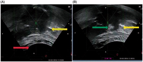
Figure 2. Contrast-enhanced MR images from a 36 years old patient with multiple uterine fibroids. (A). A sagittal view image obtained before HIFU treatment showed the size of uterus was 9.6 × 7.6 × 7.0 cm3, multiple uterine fibroids were detected (arrows). (B). A sagittal view image obtained one day after HIFU showed the fibroids were completely ablated (arrows). (C). A sagittal view image obtained 6 months after HIFU showed the size of uterus was 8.0 × 7.3 × 6.4 cm3, the rate of fibroid volume reduction was 89.6% (arrows).
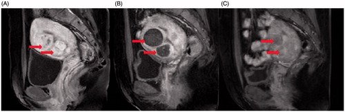
Table 1. Baseline characteristics of patients with multiple uterine fibroids.
Table 2. USgHIFU treatment results of patients with multiple uterine fibroids.
Table 3. Side effects and complications during or after the procedure.
Table 4. Comparison of UFS and QOL pre-HIFU and post-HIFU.
Table 5. Shrinkage rate of 120 fibroids in 21 followed to 6 months during follow-up interval.