
Figures & data
Table 1. Demographic and clinical characteristics before radiofrequency ablation (RFA) of primary thyroid cancer.
Figure 1. Radiofrequency ablation (RFA) of anaplastic thyroid carcinoma (A) Initial computed tomography (CT) image of an 80-year-old woman revealed 8 cm mass in the left thyroid gland with neck bulging and trachea deviation, proven as anaplastic thyroid carcinoma on core-needle biopsy. (B) Follow-up CT after first RFA (two days after initial RFA) revealed a 8.5 cm ablation zone with decreased enhanced portion of the mass. (C) Follow-up CT after second RFA (five days after initial RFA) revealed a 8.5 cm ablation zone. (D) On last follow-up CT after third RFA (39 days after initial RFA), CT revealed a 10 cm mass in the thyroid gland with unchanged neck bulging and trachea deviation.
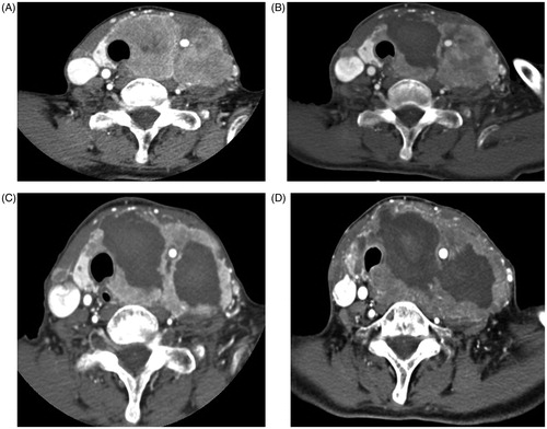
Figure 2. Gradual reduction of papillary thyroid macrocarcinoma after two sessions of RFA (A) Initial ultrasonography (US) of a 77-year-old woman revealed 8.0 cm predominantly cystic mass in the left thyroid gland. (B) After first RFA, US revealed a smaller but persistent, 2.4 cm ill-defined ablation zone in the left thyroid gland. (C) After second RFA, US revealed a smaller, 1.9 cm ill-defined ablation zone in the left thyroid gland. (D) At 12-months follow-up after two sessions of RFA, US revealed a smaller, 1.7 cm ill-defined ablation zone in the left thyroid gland. (E) At 18-months follow-up, US revealed a smaller with decreased calcification, 1.4 cm ill-defined ablation zone in the left thyroid gland.
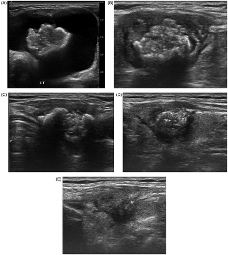
Figure 3. Gradual reduction and complete disappearance of papillary thyroid microcarcinoma after one session of radiofrequency ablation. (A) Initial US of a 49-year-old woman revealed a 0.8 cm mass in the left thyroid gland proven as papillary thyroid carcinoma on CNB. (B) At one-year follow-up after one session of RFA, US revealed a smaller, 0.4-cm ill-defined ablation zone in the left thyroid gland. (C) At 24-months follow-up after RFA, US revealed complete disappearance of ablation zone with subtle scar in the left thyroid.
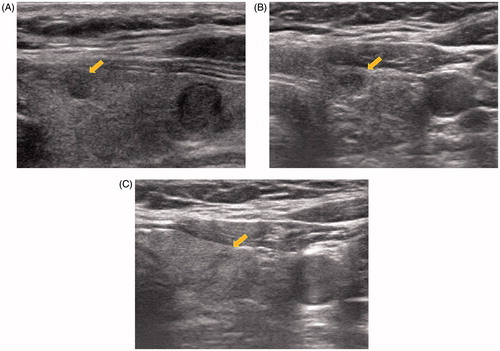
Table 2. Outcome of radiofrequency ablation with primary thyroid cancerTable Footnotea.
Table 3. Treatment parameters of primary thyroid cancer.