
Figures & data
Figure 1. Pre-operative and post-operative MRI. T2-weighted images of a 51-year-old woman with uterine fibroids but no abdominal wall scar. (A) Axial view before HIFU treatment, indicating no abnormal signals in the abdominal wall; (B) Hyperintense signals on the axial view after HIFU treatment (indicated by the arrowhead); T2-weighted images and contrast-enhanced MRI images of a 41-year-old case with uterine fibroids and an abdominal wall scar. (C) Sagittal view before HIFU treatment, indicating no abnormal signals in the abdominal wall; (D) Hyperintense signals in the sagittal view after HIFU treatment (indicated by the arrowhead); (E) Sagittal contrast-enhanced image before HIFU treatment; (F) Sagittal contrast-enhanced image after HIFU treatment, uterine fibroids located in the anterior wall and posterior wall of the uterus were almost completely ablated with non-perfusion area in the fibroids (indicated by the arrowhead).
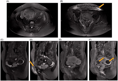
Table 1. Patient baseline characteristics.
Table 2. Treatment results of HIFU for patients.
Table 3. Univariable logistic regression analysis of variables associated with thermal damage of the abdominal wall structures.
Table 4. Multiple logistic regression analysis of variables associated with thermal damage of the abdominal wall structures.
Table 5. Post-operative adverse reactions of patients [n (%)].