Figures & data
Figure 1. The illustration of two types of complete MWA: concentric type and nonconcentric type. The largest ablated margin (L) around the tumour was measured in US. When the largest margin was <10 mm, the case was defined as accurate ablation.
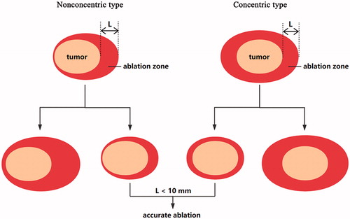
Figure 2. Macroscopic appearance of excised specimen after surgery (a) and HE staining (100×) of ablative area (b) and surrounding tissue (c) in 42-year-old woman. (a) The ablated area (arrow) is easily identified. (b) The ablated tumour is replaced by the stroma, and seldom nucleus is observed. (c) The surrounding tissue of the ablated tumour shows inflammatory cells infiltration.
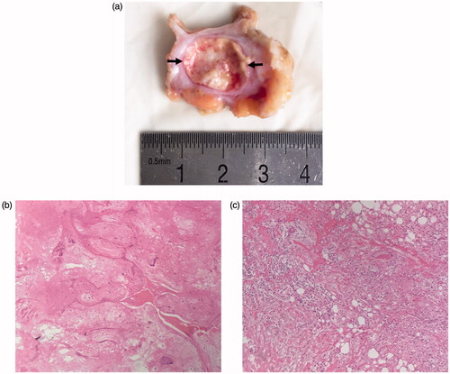
Figure 3. Conventional US and CEUS images in a 38-year-old woman before and 3 days after MWA. (a) Conventional US shows a tumour with a clear margin before MWA. (b) CEUS shows heterogeneous enhancement before MWA. (c) The tumour (arrow) shrank after ablation, but it is still clear in US. (d) The tumour (arrow) locates in the centre of the ablation zone (arrow-head) in CEUS. This case is defined as concentric type.
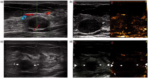
Figure 4. Nonconcentric type of ablation in a 26-year-old woman. (a) A clear tumour (arrow) in US is observed before MWA. (b) Enhancement of the tumour is observed in CEUS before MWA. (c) The shrank tumour (arrow) is still clear in US Week 1 after ablation. (d) The tumour (arrow) does not locate in the centre of the ablation zone (arrow-head) in CEUS.
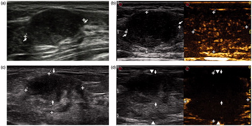
Figure 5. The largest margin (a) and cumulative probability of accurate ablation (b) of these sequential 52 cases. All seven nonaccurate ablations are in the training group (the first 30 cases). In the practiced group (the last 22 cases), the cumulative probability steadily increases.
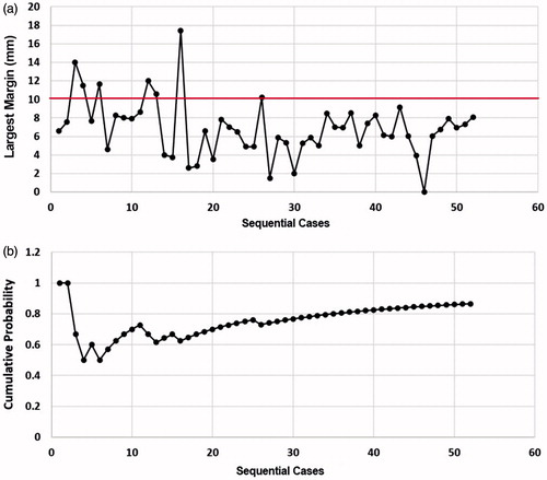
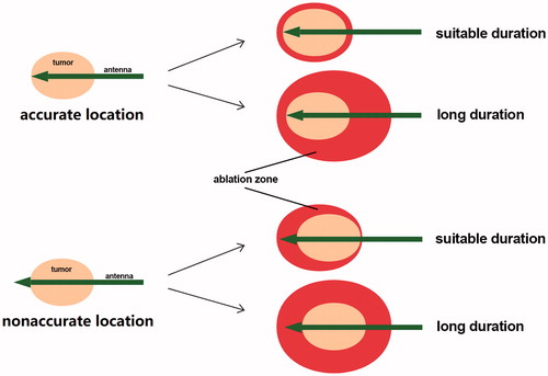
Figure 6. The role of the antenna location and the ablation duration in accurate MWA of benign breast tumours. For complete ablation, four major cases may occur. Both accurate location of the antenna and suitable duration contribute to ideal MWA in the treatment of benign breast tumours.