Figures & data
Figure 1. RFA thermal dose settings during hepatic ablation stimulate variable distant subcutaneous tumor growth. Animals implanted with subcutaneous R3230 tumors were treated with low- (60 °C × 10 min), medium- (70 °C × 5 min) and high-dose (90 °C × 2 min) hepatic RF ablation compared to sham treatment. After hepatic ablation (Day 0), the growth rates of the distant tumors significantly increased for low- and medium-dose RFA compared to high-dose RFA and sham treatment. This resulted in significantly larger tumor size (p < .05) at day 7 for low- and medium-dose RFA compared high-dose RFA and sham treatment. A significant increase in tumor growth was still seen in high-dose RFA compared to sham (p < .05); however, the effect is much less pronounced.
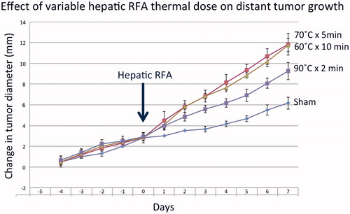
Table 1. Summary of subcutaneous tumor growth and proliferative index for different hepatic RF ablation parameters without and with adjuvant micellar quercetin. A detailed summary of tumor growth data (including pre- and post-treatment tumor measurements growth curve slopes) is provided. Immunohistochemistry quantification of proliferative index (Ki-67% cell positivity) is also provided. Statistically significant differences (defined as p < .05) are annotated.
Table 2. Summary of changes in serum, liver, and distant tumor HGF and VEGF levels for variable hepatic RF ablation without and with adjuvant micellar quercetin therapies.
Figure 2. Adjuvant micellar quercetin administered at the time of hepatic RFA suppresses distant subcutaneous tumor growth. Animals implanted with subcutaneous R3230 tumors were treated with low-dose (60 °C × 10 min) and high-dose (60 °C × 10 min) hepatic RFA with and without single-dose micellar quercetin (Mic-Qu) compared to sham treatment and micellar quercetin alone (total 6 treatment arms). After hepatic ablation (Day 0), the growth rates of the distant R3230 tumors significantly increased for low-dose RFA and to a lesser degree, high-dose RFA compared to sham treatment. Adjuvant micellar quercetin significantly reduced distant tumor growth rates of both doses down to near-baseline levels (p < .01 for all comparisons).
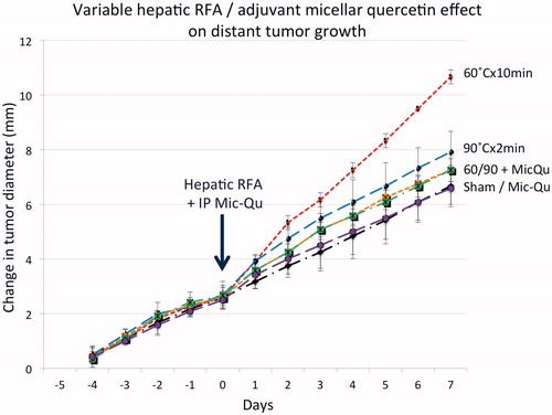
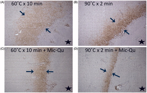
Figure 3. Adjuvant micellar quercetin suppresses HSP expression in the periablational rim. Marked hepatic ablation-induced HSP70 expression (bracketed by arrows in all images) after (A) low-dose (60 °C × 10 min) and (B) high-dose (90 °C × 2 min) ablation is significantly reduced with adjuvant single-dose micellar quercetin administered at the time of ablation (Day 0) for both (C) low- and (D) high-dose RFA (p < .01 for all comparisons). The black star denotes the ablation zone.