
Figures & data
Table 2. Demographic and clinical characteristics of patients.
Table 3. Tumor, ablation, and complication characteristics of 25 lesions in 23 patients with IPA.
Table 4. Symptoms and laboratory and radiological findings of 23 patients with IPA.
Table 5. Diagnosis, antifungal therapy, and outcomes in 23 patients with IPA after lung MWA.
Figure 1. (a) A contrast-enhanced CT scan showing a single neoplasm, 3.5 × 2.4 cm, adjacent to mediastinal pleural in the right upper lobe before MWA. (b) MWA was performed at a power of 70 W for an accumulated total time of 11 min. (c) A follow-up CT scan at 24 hours after MWA showing a GGO-like reaction band around the lesion that almost surrounded the entire tumor. (d) At 28 days after MWA, the ablation zone was replaced by a large uneven thick-walled cavity containing a mass of irregular consolidation surrounded by patchy infiltration. (e) A chest CT scan at 6 weeks after MWA showing shrinkage of the cavity, incrassation of the wall, and aggravated infiltration. (f) A chest CT scan at 6 weeks after MWA also showing multiple patchy infiltrations and nodules scattered in both lung fields.
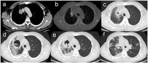
Figure 2. (a) A chest CT image showing a primary tumor 4.5 cm in diameter abutting the thoracic aorta and left lower pulmonary artery in the superior segment of the left lower lobe. (b) Despite a mild pneumothorax, MWA was successfully completed with a power of 70 W for a total of 21 min. (c) A CT scan 3 weeks after MWA showing a thin-walled cavity with an irregular luminal surface in the ablation zone. (d) The same CT scan 3 weeks after MWA showing coexistence of the cavity and nodules with a halo sign in the left upper lobe. (e) The same CT scan 3 weeks after MWA showing consolidation with cavitation in the right upper lobe and an uneven think-walled cavity containing pedunculated contents in the left lower lobe. (f) A CT scan 5 weeks after MWA showing diffuse consolidations with cavitation, infiltrations and nodules in bilateral lungs. (g) The CT scan 5 weeks after MWA showing the cavity emptying inside, as well as nodules enlarged and cavitated in the left upper lobe.
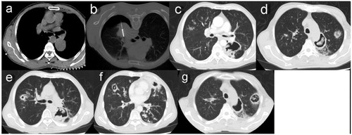
Table 1. Criteria for proven and probable invasive pulmonary aspergillosis.