
Figures & data
Figure 1. A 51-year-old male. (a, b) Contrast-enhanced CT indicates a liver tumor located in segment 7. Arterial enhancement and delay wash-out were observed. (c) A hypoechoic lesion in US. (d) Real-time US was successfully matched with immediately acquired preablation 3DUS. (e) Real-time US was matched with preablation CT volume images successfully. (f) Ablation was applied under the guidance and monitoring of US. (g) CEUS was performed and fused with preablation 3DUS. The ablation zone completely covered the target tumor and the 5-mm ablative margin. (h) CEUS fused with preablation CT indicated the ablation zone completely covered the target tumor and the 5-mm ablative margin. (i) Subsequent contrast-enhanced CT within three months confirmed that the tumor had been completely ablated.
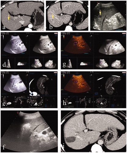
Figure 2. A 43-year-old male. (a, b) Contrast-enhanced CT indicated a massive hepatocellular carcinoma in the left liver with a small lesion located in segment 8. (c) Hepatectomy was performed before ablation, and the small lesion was hypoechoic on US. (d) Deformation of liver after hepatectomy led to the failure of registration of US and preablation CT volume images. (e) Immediately acquired 3DUS volume images were successfully matched to the real-time US. (f) After the ablation procedure, CEUS was performed and fused with preablation 3DUS. The ablation zone completely covered the target tumor and the 5-mm ablative margin. (g) Subsequent contrast-enhanced CT within three months confirmed that the tumor had been completely ablated.
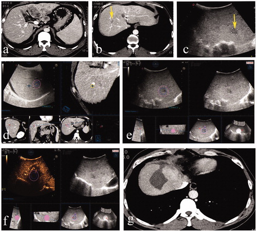
Figure 3. A 46-year-old male. (a,b) Contrast-enhanced MRI indicated a small recurrent tumor located in segment 4. (c) The lesion was inconspicuous in the approximated location on US. (d,e) Arterial enhancement and delay wash-out in Contrast-enhanced US was observed. (f) Real-time US was matched with preablation MRI volume images successfully. (g) Ablation was performed. (h) CEUS fused with preablation MRI indicated the ablation zone completely covered the target tumor and the 5-mm ablative margin. (i) Subsequent contrast-enhanced CT within three months confirmed that the tumor had been completely ablated.
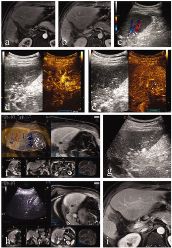
Table 1. Baseline features of the study population.
Table 2. The applicable rate and success rate of registration in CT/MR-CEUS and 3DUS-CEUS fusion imaging techniques.
Table 3. Causes for inapplicable CT/MRI-CEUS and US-CEUS fusion imaging.
Table 4. Causes for the failure of registration in CT/MRI-CEUS and US-CEUS fusion imaging techniques.
Table 5. Influence of combined operations or procedures during the ablation procedure on the success rate of registration in two fusion imaging techniques.
Table 6. Influence of different combined operations or procedures during ablation procedure on success rate of registration in two fusion imaging techniques.