
Figures & data
Figure 1. (Top image) Overview of STS evaluation and critical structure measurements in the near field. Each STS was evaluated in three planes. This image illustrates an axial projection (CT image) of an STS tumour present in the right flank. The target cell (4 mm) is positioned centrally within the constructed PTV, and the HIFU beam is aligned to the target cell. Measurements included (A) the closest distance from the skin to any tumour margin, with (B) the distance from the skin to the centre of the tumour in the near-field, and (C) the distance from the skin to the deepest tumour margin in the far field, both acquired along the HIFU beam path. (Bottom image) Overview of STS evaluation and critical structure measurements in the far field. Dorsal projection of the same tumour in the right flank. The target cell is unchanged in location and remains the same point of reference for measurements along the beam axis. Critical structure measurements for bone were acquired from (A) the centre of the tumour to nearest bone structure in the far field and (B) the tumour margin to nearest bone structure in the far field.
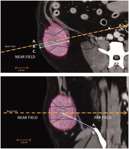
Table 1. Total number and relative percentage of canine STS by both tumour type and tumour site.
Figure 2. Anatomic distribution of targetable STS tumours by body regions. Pie charts placed at sites labelled head, truncal/axillary, spine/paraspinal, appendicular and abdomen/disseminated illustrate the distribution of all soft tissue sarcomas (53) as, ≥50% targetable (green), <50% targetable (yellow) and non-targetable (red) tumour categories. The spine/paraspinal and head sites had the largest relative percentage of non-targetable STS. Image adapted with permission [Citation55].
![Figure 2. Anatomic distribution of targetable STS tumours by body regions. Pie charts placed at sites labelled head, truncal/axillary, spine/paraspinal, appendicular and abdomen/disseminated illustrate the distribution of all soft tissue sarcomas (53) as, ≥50% targetable (green), <50% targetable (yellow) and non-targetable (red) tumour categories. The spine/paraspinal and head sites had the largest relative percentage of non-targetable STS. Image adapted with permission [Citation55].](/cms/asset/1ccbcd70-30ca-44de-94f7-19813fbb7294/ihyt_a_1489072_f0002_c.jpg)
Figure 3. Distribution of STS tumour types with the relative percentage of targetability. STS tumour types that are ≥50% targetable are depicted in green and STS tumours <50% targetable are depicted in yellow, while STS deemed non-targetable are illustrated in red. PNST had the largest relative percentage of tumours that were not targetable followed by anaplastic sarcoma, unclassified soft tissue sarcoma and fibrosarcoma.
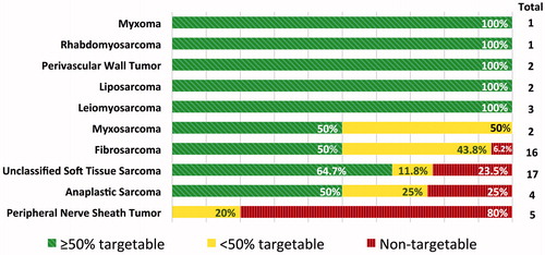
Figure 4. Representative examples of canine STS at seven anatomic sites. The PTV was selected as an ellipsoid (purple). A 4 mm diameter treatment cell (green/blue) and the resulting approximate outline of the HIFU beam (yellow) were aligned with the centre of the PTV to optimise the treatment window based on tumour location, and the patient was positioned to allow the greatest unobstructed access to the target. The orange rectangle around the target represents a safety zone in which high temperatures can occur. (A) Head: ≥50% targetable, left maxillary fibrosarcoma with a portion of the tumour invading the infraorbital foramen and left orbit (green arrow); (B) head: <50% targetable, unclassified STS of the rostral right mandible with geographic bone lysis; (C) head: not targetable, unclassified STS within the right nasal cavity; (D) truncal: ≥50% targetable, subcutaneous unclassified STS dorsal to the pelvis; (E) truncal: <50% targetable, unclassified STS medial to the left scapula; (F) appendicular: ≥50% targetable, subcutaneous anaplastic sarcoma of the left distolateral humerus; (G) spine: ≥50% targetable, unclassified STS in the right cervical region with invasion of the spinal canal at C6 (green arrow); (H) spine: <50% targetable, left paraspinal myxosarcoma involving ribs 10, 11 and 12 and invasion of the spinal canal at T10–T11 resulting in extradural spinal cord compression (green arrow); (I) spine: Not targetable, PNST involving the right spinal nerve segment at C4–C5 with extension to the spinal cord (green arrow); (J) abdominal: ≥50% targetable, extramural small intestinal fibrosarcoma; the ellipsoid ROI approximates the PTV, however, additional targets could be selected during therapy to achieve more conformal treatment; (K) axillary: ≥50% targetable, left axillary myxosarcoma partially incorporating the left first rib (green arrow); and (L) axillary: <50% targetable, left axillary PNST.
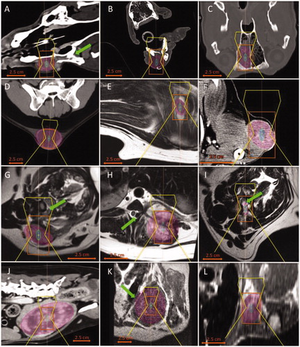
Table 2. Total number and relative percentage of targetable canine STS by both tumour type and tumour site.
Table 3. Number and relative percentage of STS for both total tumour volume (TTV) and planned treatment volume (PTV) that met the criteria for optimal treatment volume (≤200 cm3), optimal target depth (≤8 cm), maximum target depth (≤11 cm), or met both criteria for optimal volume and optimal target depth (≤200 cm3 and ≤8 cm), and optimal treatment volume and maximum target depth (≤200 cm3 and ≤11 cm).
Table 4. Contingency coefficients summary for the geometric/anatomical constraints imposed by nearby critical structures when compared by STS tumour type, tumour site and targetability.