Figures & data
Figure 1. US scans in a 57-year-old woman: (a) US scans showed that the largest diameter was 3.67 cm; (b) US scans showed that the two other perpendicular diameters were 2.46 cm and 2.28 cm, respectively. (c) Contrast-enhanced ultrasonography (CEUS) showed a cyst-solid mixed TN. The TN’s volume was 10.8 ml, of which the solid portion was 95.1%; the arrow highlights the central region of the TN.
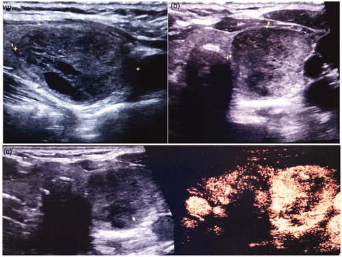
Figure 2. The same patient as in Figure 1. (a) In the MWA procedure, the ‘hydrodissection technique’ was applied. The isolation zone is marked with an arrow in the figure. (b) MWA antenna could be visualized as a hyperechoic line in the MWA process under US. The area that surrounds the antenna also shows the hyperechoic region. (c) CEUS showed that approximately 99% of the TN had been ablated.
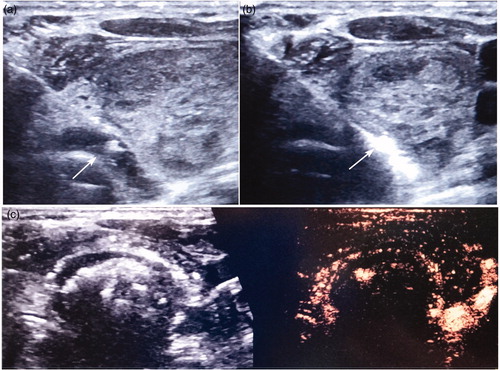
Table 1. Baseline characteristics of patients with TNs treated by US-guided PMWA and conventional thyroidectomy before and after matching.
Figure 3. (a) A 61-year-old woman in the thyroidectomy group; the picture shows her early postoperative wound within a highly visible area; (b) The same patient as in Figures 1 and 2, the picture shows her cosmetic results of US-guided PMWA, where there is no obvious cervical scar in her neck.
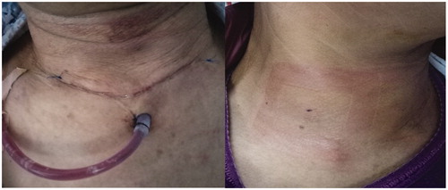
Table 2. Operation results in the MWA and conventional thyroidectomy groups in a propensity-matched population of patients with TNs.
Table 3. Serum Concentrations of CRP in the MWA and conventional thyroidectomy groups in a propensity-matched population of patients with TNs (mg/L).
Table 4. Serum Concentrations of IL-6 in the MWA and conventional thyroidectomy groups in a propensity-matched population of patients with TNs (ng/L).
Table 5. Serum Concentrations of TNF-α in the MWA and conventional thyroidectomy groups in a propensity-matched population of patients with TNs (mg/L).
Table 6. Treatment results in the MWA group in a propensity-matched population of patients with TNs.
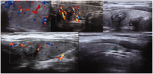
Figure 4. US scans in a 30-year-old woman: (a) US reveals a cyst-solid mixed TN. The TN’s volume was 4.1 ml, of which the solid portion was 83.6%. (b) One month after the ablation, the volume of the TN decreased to 2.5 ml with a VRR of about 39.6%. (c) Three months after US-guided PMWA, the volume was 1.2 ml, with a VRR of about 73.8%. (d) Twelve months after US-guided PMWA, the volume was 0.03 ml with a VRR of about 97.6%.