Figures & data
Table 1. Information on the 12 cases of patients with giant hemangiomas.
Figure 1. Needle placement description.
Table 2. Discomfort and complications after MWA for 13 giant hemangiomas.
Table 3. Liver function changes after ablation.
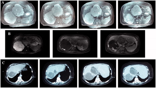
Figure 2. A 67-year-old female with a giant hepatic hemangioma of 13 cm in diameter. (A) Preoperative MRI (size: 13 cm × 9 cm). (B) MRI after the first ablation (size: 8 cm × 7 cm). (C) MRI after the second ablation (size: 10.8 cm × 7.8 cm).
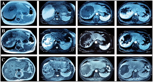
Figure 3. A 37-year-old male with two giant hepatic hemangiomas of 12.5 and 11.7 cm in diameter. (A) Preoperative MRI (size: 12.5 cm × 10.0 cm, 11.7 cm × 9.3 cm). (B) MRI after the first ablation (size: 7.5 cm ×6.2 cm). (C) MRI after the second ablation (size: 7.1 cm × 6.4 cm).