
Figures & data
Figure 1. Flow chart.
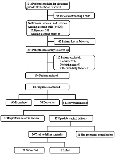
Table 1. Baseline characteristics of 81 pregnant women at the time of ultrasound-guided HIFU ablation treatment.
Figure 2. Images for a 28-year-old woman with two vaginal deliveries after ultrasound-guided HIFU ablation treatment for a fibroid. (a) Contrast-enhanced ultrasound (CEUS) obtained before treatment showed enhancement of the fibroid (white arrow) on the posterior wall of the uterus. (b) Immediate CEUS obtained after treatment showed a non-enhanced region (53.7 cm3). The margins of the non-enhanced region (white arrow) were shown at the ablated region. (c) and (d) Sagittal view of MRI obtained before treatment showed enhancement of the fibroid (red arrow) on the posterior wall of the uterus. (e) and (f) Sagittal view of an MRI obtained 3 months after ultrasound-guided HIFU ablation showed a decreased size of the fibroid (red arrow). The volume of the fibroid was 35.5 cm3. The woman was pregnant after 6 months and 24 months following treatment and gave birth to two healthy babies weighing 3.3 kg and 3.0 kg, respectively.
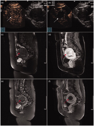
Table 2. Clinical characteristics and obstetric outcomes of 19 women achieving successful vaginal delivery after ultrasound-guided HIFU ablation treatment with summary statistics.
Table 3. Clinical characteristics and obstetric outcomes of 5 women who experienced failed vaginal delivery after ultrasound-guided HIFU ablation treatment with summary statistics.