
Figures & data
Table 1. Patient characteristics.
Figure 1. Ultrasound and contrast-enhanced ultrasound imaging (from the guiding ultrasound device of the high-intensity focussed ultrasound[HIFU] unit) in a 36-year old woman with abdominal wall endometriosis (AWE). (A) Before HIFU, a hypoechoic nodule (25 × 20 mm) between the subcutaneous fat layer and rectus abdominis (distance from skin surface, 5 mm) (white arrow). (B) Before HIFU, the nodule was enhanced oncontrast-enhanced ultrasoundafter contrast agent injection (white arrow). (C) One day after HIFU, the nodule was ablated with complete absence of perfusion (white arrow), and cyclic pain disappeared. The nodule disappeared and no recurrence occurred during the 3-year follow-up.
![Figure 1. Ultrasound and contrast-enhanced ultrasound imaging (from the guiding ultrasound device of the high-intensity focussed ultrasound[HIFU] unit) in a 36-year old woman with abdominal wall endometriosis (AWE). (A) Before HIFU, a hypoechoic nodule (25 × 20 mm) between the subcutaneous fat layer and rectus abdominis (distance from skin surface, 5 mm) (white arrow). (B) Before HIFU, the nodule was enhanced oncontrast-enhanced ultrasoundafter contrast agent injection (white arrow). (C) One day after HIFU, the nodule was ablated with complete absence of perfusion (white arrow), and cyclic pain disappeared. The nodule disappeared and no recurrence occurred during the 3-year follow-up.](/cms/asset/2c392334-b7c6-41c7-b352-6515e6b27c21/ihyt_a_1511836_f0001_c.jpg)
Figure 2. Ultrasound and skin imaging in a 35-year old woman with endometriosis of the abdominal muscle layer. (A) Before HIFU, sagittal guided ultrasound showed a low echogenic nodule of approximately 33 mm in the right subcutaneous muscle layer of the abdominal wall incision. Distances from the superficial and deep areas of the lesion to skin surface were 6 mm and 29 mm, respectively. (B) The bladder was filled with 400 ml normal saline to push the intestine behind the lesion. During the operation, ultrasound energy of fixed-point emission was 12000 J, and the focus position showed a massive hyperechoic change. There was no significant echo change in the superficial tissue of the lesion. (C) After HIFU, skin surface was slightly swollen and the skin was intact.
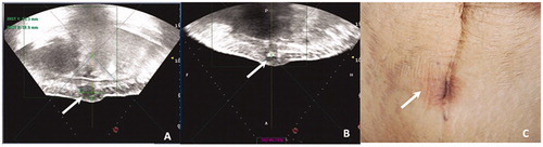
Table 2. Technical success rates and medical cost.
Table 3. Adverse events in the surgery and HIFU groups.
Table 4. Clinical evaluation in the surgery and HIFU groups.