
Figures & data
Table 1. Patients baseline characteristics and treatment data.
Figure 1. Contrast enhanced magnetic resonance imaging (CE-MRI) before and after treatment: (A1) anterior wall and mildly enhanced uterine fibroids; (B1) lateral wall and moderately enhanced uterine fibroids; (C1) posterior wall and significantly enhanced uterine fibroids; CE-MRI after treatment shows that the ablation area is not enhanced (A2, B2 and C2).
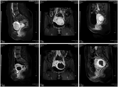
Table 2. The multivariable regression model and analysis of varianceTable Footnotei.
Table 3. Coefficient of multivariable regression modelTable Footnotea of NPVR.
Table 4. The multivariable regression model and analysis of varianceTable Footnoteg.
Table 5. Coefficient of multivariable regression modelTable Footnotea of EEF.