Figures & data
Figure 1. PD-L1 and CD8+ TIL expression (40×) (A, PD-L1-positive expression; B, PD-L1-negative expression; C, CD8+ TIL and D, CD8– TIL).
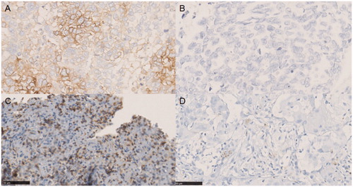
Figure 2. The patients enrolled in the study.
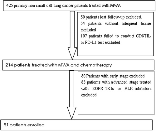
Table 1. The baseline characteristics of enrolled patients
Table 2. Correlation between PD-L1 expression, CD8+ TIL expression and clinicopathological characteristics.
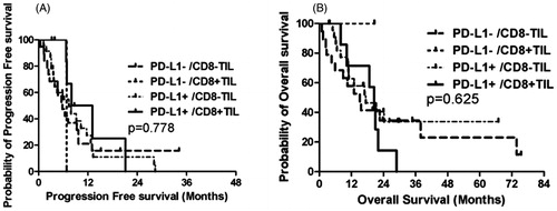
Figure 3. Kaplan–Meier plots of PFS and OS. (A) PFS and PD-L1 expression; (B) OS and PD-L1 expression; (C) PFS and CD8+ TIL and (D) OS and CD8+ TIL.
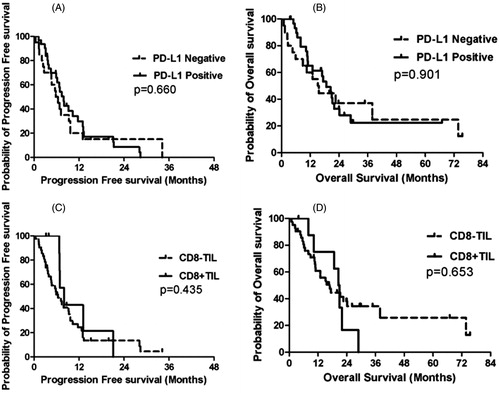
Table 3. Correlation between PD-L1 expression, CD8+ TIL expression and survival.
Figure 4. Kaplan–Meier plots of PFS and OS. (A) PFS and PD-L1 expression and CD8+ TIL and (B) PFS and PD-L1 expression and CD8+ TIL.