Figures & data
Table 1. Univariate analysis to evaluate the relationship between the sacral injury and the features of fibroids.
Table 2. Multivariable binary logistic regression analysis to evaluate the correlation of sacral injury with the significant factors of univariate analysis.
Table 3. Evaluation of the relationship between sacral injury and treatment parameters according to univariate analysis.
Table 4. corralation of the sacral injury with the significant treatment parameters according to multivariate analysis.
Table 5. Summary of postprocedural adverse effects.
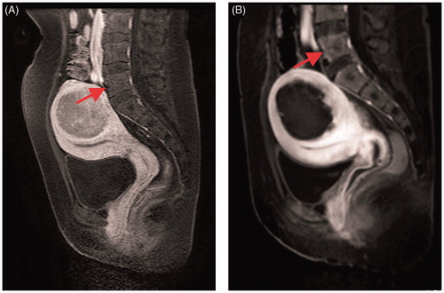
Figure 1. CE-MRI before and after USgHIFU ablation: (A) sacrum shows normal before treatment; (B) sacrum shows non-perfused upon CE-MRI immediately after treatment.
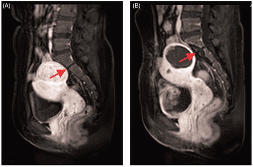
Figure 2. CE-MRI before and after USgHIFU ablation: (A) the fifth lumbar spine shows normal before treatment; (B) the front edge of the fifth lumbar spine shows non-perfused upon CE-MRI immediately after treatment.