
Figures & data
Table 1. Characteristics of Patient.
Figure 1. Overall survival of patients.
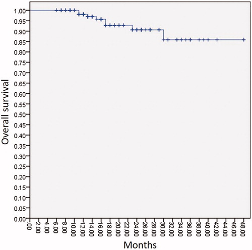
Figure 2. Local tumor progression based on per patient and per lesion.
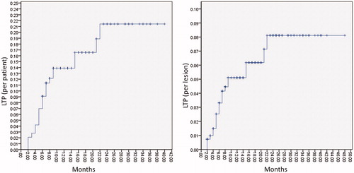
Figure 3. A 51-year-old female patient with four CRLM. (A) After artificial hydrothorax injection of 600 ml of saline (*), percutaneous US clearly showed one lesion 11 mm in diameter (thick arrow) under the diaphragm. (B) CEUS showed the lesion (thick arrow) with homogeneous hyper-enhancement in the arterial phase (30 s). (C) The lesion (thick arrow) presented hypo-enhancement in the late phase (180 s). (D) The antenna (thin arrow) was inserted into the lesion 5 mm beyond the deep margin of the lesion (thick arrow) under US guidance, and then the MWA procedure was initiated (50 W, 10 min). (E) US showed that the lesion (thick arrow) was completely covered by hyperechoic gas during the ablation procedure. (F–G) The ablation zone (thick arrow) showed non-enhancement in the arterial and venous phases of CEMRI 1 month after ablation. (H–I) The ablation zone (thick arrow) was smaller and showed non-enhancement in the arterial and venous phases of CECT 30 months after ablation.
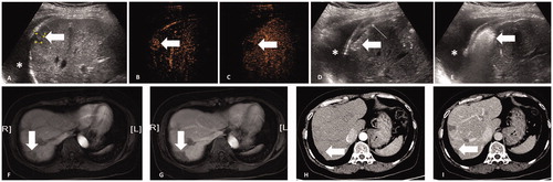
Table 2. Factors associated with local tumor progression.
Table 3. Factors to predict local progression.
Table 4. Major and minor complications of MWA.
Table 5. The degree of pain after MWA.