Figures & data
Table 1. The baseline characteristics of the study population.
Figure 1. A 55-year-old male. (a, b, c) Contrast-enhanced ultrasound and MRI indicated a small liver tumor located in segment 5 abutting the gallbladder with a maximum diameter of 14 mm. (d, e) The electrode was inserted parallel to the gallbladder wall with a minimum distance of 10 mm. (f) Contrast-enhanced ultrasound demonstrated a complete ablation and intact perfusion of gallbladder wall. (g) Contrast-enhanced MRI one month after the ablation procedure confirmed the technical efficacy. (h) Normal gallbladder wall was showed in the follow-up period.
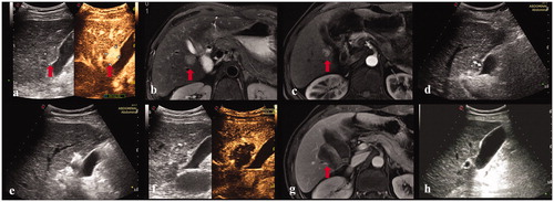
Figure 2. A 28-year-old female. (a, b) Contrast-enhanced ultrasound and MRI indicated a liver tumor located in segment 5 abutting the gallbladder with a maximum diameter of 27 mm. The thickness of gallbladder wall was 4 mm. (c) The electrode was inserted parallel to the gallbladder wall with a minimum distance of 8 mm. (d) The observation of gallbladder wall was affected by the gas produced by ablation. (e) Contrast-enhanced ultrasound showed that the perfusion of gallbladder wall was intact and the index tumor had been completely ablated. (f) Three day after ablation, the thickness of gallbladder wall increased to 9 mm. (g) Contrast-enhanced MRI one month after the ablation procedure confirmed the complete ablation of the index tumor. (h) Four months after ablation, the thickness of gallbladder wall was restored to 4 mm.
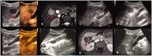
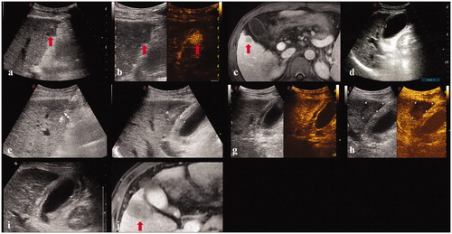
Figure 3. A 45-year-old male. (a, b, c). Contrast-enhanced ultrasound and MRI indicated a liver tumor located in segment 5 adjacent to the gallbladder with a maximum diameter of 18 mm. (d) Before ablation, the thickness of gallbladder wall was 5 mm. (e) The electrode was inserted parallel to the gallbladder wall with a minimum distance of 6 mm. (f) The thickness of gallbladder wall increased to 10 mm immediately after ablation. (g, h). Contrast-enhanced ultrasound showed that the perfusion of gallbladder wall was intact and the index tumor had been completely ablated. (i) The thickness of gallbladder wall was 9 mm five days after ablation. (j) Contrast-enhanced MRI one month after the ablation procedure confirmed the technical efficacy and the thickness of gallbladder wall was restored to 5 mm.