
Figures & data
Figure 1. Flow diagram showing patient selection criteria.
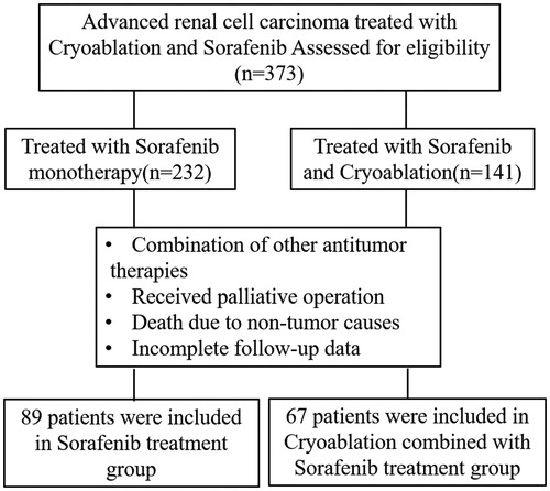
Table 1. Clinical data of 156 patients with advanced renal cell carcinoma.
Figure 2. Progression-free survival of patients in the cryoablation + sorafenib and sorafenib-only groups.
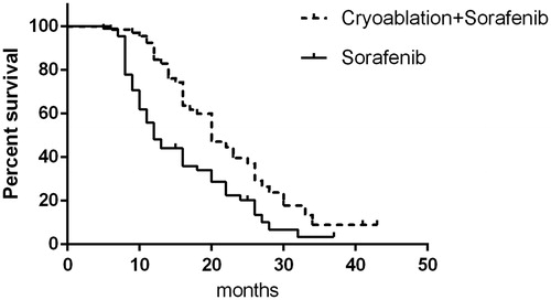
Figure 3. Overall survival of patients in the cryoablation + sorafenib and sorafenib-only groups.
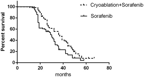
Figure 4. Comparison of progression-free survival between complete cryoablation and incomplete cryoablation.
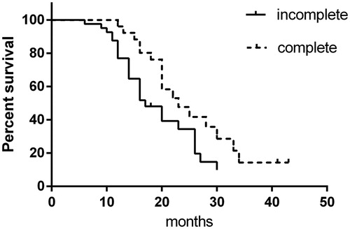
Figure 5. Comparison of overall survival between complete cryoablation and incomplete cryoablation.
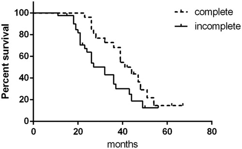
Figure 6. A 60-year-old man with a massive tumor on the right kidney accompanied by multiple (lung and bone) metastases who underwent cryoablation combined with sorafenib. (A, B) Preoperative CT scans show that the tumor was located in the subrenal pole and involved the renal hilum, with the largest tumor diameter of 6.8 cm. (C, D) CT scans show incomplete ablation of the renal hilum tumor during cryoablation. (E, F) CT scan at 1 month after cryoablation shows that most of the tumor body is completely necrotic and that there is residual tumor tissue in the renal hilum.
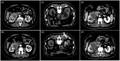
Figure 7. A 48-year-old woman with a tumor on the left kidney accompanied by bone metastasis who underwent cryoablation combined with sorafenib. (A, B) Preoperative CT scans show that the tumor was located in the lower pole of the left kidney, with the largest tumor diameter of 7 cm. (C, D) CT scans show complete ablation of the tumor during cryoablation. (E, F) CT scan at 1 month after cryoablation shows that the tumor is completely necrotic.
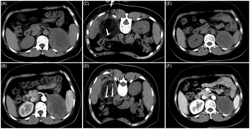
Figure 8. Forest plot for subgroup analysis of OS.
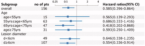
Table 2. Clinical efficacy evaluation.
Table 3. Immune-related indicators before and after treatment in each group.
Table 4. Hepatic and renal function-related indicators before and after treatment in each group (mean ± s).
Table 5. Adverse events in cryoablation + sorafenib and sorafenib-only groups.