Figures & data
Table 1. Baseline clinical characteristics of RT patients with SHPT treated with MWA.
Table 2. Laboratory data of RT patients with SHPT before or after MWA.
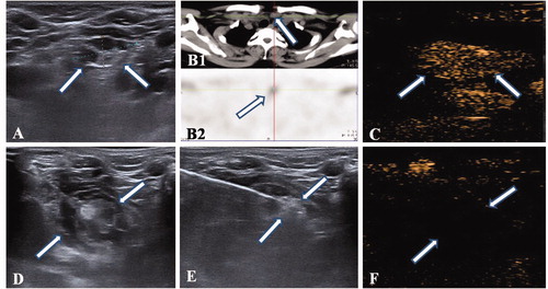
Figure 1. MWA of SHPT nodule in a patient who had undergone successfully renal transplantation. (A) The nonuniform hypoechoic nodule (Arrow) was disclosed by preablation ultrasound in neck. (B1) The CT scan showed there is a small nodule (Arrow) locating in the right front of trache, which corresponds to the one in ultrasound; (B2) The nodule has radioactivity concentration (Arrow) in late phase of MIBI scan. (C) An uniform hyperenhancement of nodule (Arrow) was displayed in CEUS preablation. (D) The spacer fluid was injected surrounding the nodule, which displayed non echo area (Arrow). (E)The hyperechoic emerging inside nodule (Arrow) during ablation. (F) A non-enhancement area covered the nodule (Arrow) after ablation, which suggested a complete ablation.