Figures & data
Figure 1. Result of organs’ segmentations: 2D contours in the axial, sagittal and coronal views and 3D reconstructions.
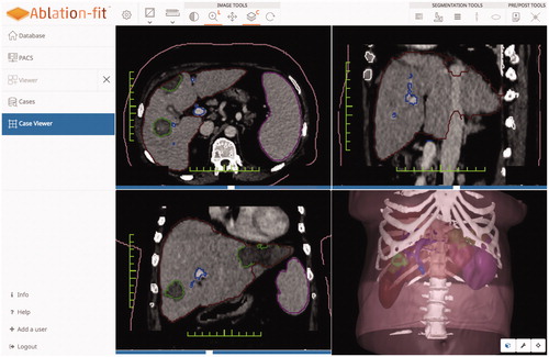
Figure 2. Categorization of post-ablation outcomes. Top left: Tumor (inner part) entirely included in the necrosis (outer part). Top right: Tumor (inner part) and 5 mm safety margin (middle part) entirely included in the necrosis (outer part). Bottom left: Tumor (inner part) entirely included in the necrosis (outer part), 5 mm safety margin (middle part) partially included in the necrosis. Arrow indicates the safety margin region not included in the necrosis. Bottom right: Tumor (inner part) and 5 mm safety margin (middle part) partially included in the necrosis (outer part). Arrows indicate the tumor region and the safety margin region not included in the necrosis.
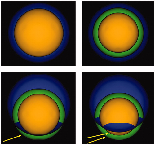
Figure 3. Result of pre- post-ablation registration: HCCs is the inner part, 5 mm margins (middle part) coagulation zone (outer part). The solid portion outside the necrosis is the unablated safety margin. Calculations rendered are: residual unablated nodule 0%, residual unablated 5 mm margin 7.8%.
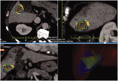
Table 1 Size of HCCs in the 1-yr follow-up CT and in the five categories of Ablation-fitTM analysis.
Figure 4. Top left: Pre-ablation HCC segmentation. Top right: Post-ablation segmentation of the necrosis volume. Bottom left: pre- post-ablation registration; the residual unablated nodule percentage is 0% and the residual unablated 5 mm margin is 12.5% (the solid portion outside the necrosis is the unablated safety margin). Bottom right: 1-year follow-up CT with necrosis segmentation showing no-LTP.
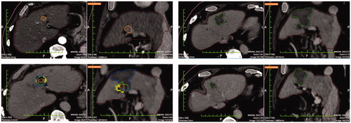
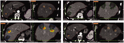
Figure 5. Top left: Pre-ablation segmentation of two HCCs in the same patient. Top right: Post-ablation segmentation of the coagulation volumes achieved. Bottom left: pre- post-ablation registration. Calculations rendered are the following: residual unablated tumor 78.9% and 100% respectively and residual unablated 5 mm margin 77.6% and 93.0% (the inner solid portion outside the necrosis is the unablated tumor and the outer solid portion outside the necrosis is the unablated safety margin). Bottom right: 1-year follow-up CT with LTPs segmentations. LTPs developed in the exact location where residual unablated tumor was shown in the pre-post-ablation registration (bottom left image).