Figures & data
Figure 1. Flowchart of the patient enrollment. RFA: radiofrequency ablation.
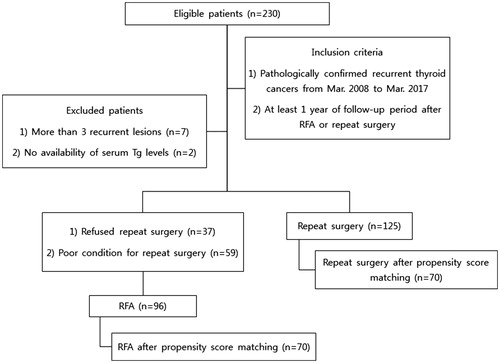
Table 1. Baseline characteristics of study population.
Table 2. Serum Tg level changes and recurrence-free survival of both groups.
Table 3. Complications after RFA and repeat surgery.
Figure 2. Ultrasound (US; a) and computed tomography (CT; c) images obtained at the initial radiofrequency ablation procedure in a 51-year old woman with recurrence at right cervical level 4 (a and c, respectivelyarrows). The recurrent lesion completely disappeared at the 98-month follow-up, as observed on US (b), and had also disappeared at the 5-month follow-up, as observed on CT (d; arrows).
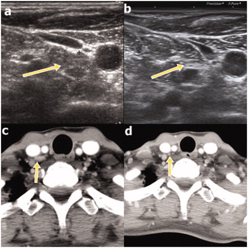
Figure 3. Ultrasound (US; a) and positron emission tomography-computed tomography (PET-CT; c) images of recurrence at right cervical level 4 in an 81-year old female patient. At the 122-month follow-up, complete disappearance of the recurrence was observed on US (b); it was also noted at the 39-month follow-up on PET-CT (d).
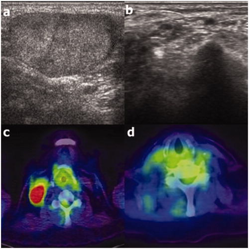
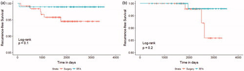
Figure 4. Kaplan–Meier recurrence-free survival curves of radiofrequency ablation (RFA; green) and surgery (red) groups before (a) and after (b) propensity score matching.