
Figures & data
Table 1. Demographic characteristics of the patients with placenta accreta.
Figure 1. Therapeutic algorithm for management of retained placenta accreta or increta with marked vascularity after abortion or delivery. After identifying vascular lesion in retained placenta tissue by transvaginal color Doppler ultrasound and MRI and serum β-hCG measurement, cases were divided into low (≤100 mIU/mL) and high (>100 mIU/mL) serum β-hCG groups. Cases with low serum β-hCG were managed by HIFU + ultrasound-guided curettage or hysteroscopic resection, while cases with high serum β-hCG were managed by HIFU + systemic MTX + ultrasound-guided curettage or ultrasound-guided hysteroscopic resection.
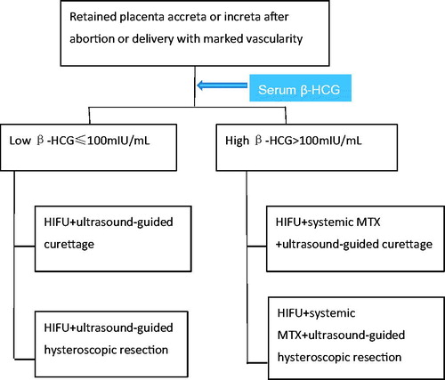
Figure 2. Retained placenta accreta marked vascularity after abortion. (A) MRI showed the retained placenta accreta as irregular low signal intensity; (B) Ultrasound before operation: 32 mm × 20 mm × 19 mm hyperechogenic shadow in the endometrial cavity with patchy blood flow signal, and unclear boundary with the muscular layer of the uterus was shown; (C) The lesion was not seen at 1 month after operation.
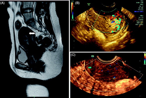
Figure 3. Retained placenta increta marked vascularity after delivery. (A) MRI showed the retained placenta increta as high signal intensity (arrow); (B) Ultrasound imaging before operation: 65 mm × 38 mm × 43 mm hyperechogenic shadow in the endometrial cavity with marked vascularity and deep myometrial invasion reached to uterine serosa was shown; (C) 25 mm × 20 mm × 23 mm hyperechogenic shadow at one month after operation; (D) 16 mm × 14 mm × 18 mm hyperechogenic shadow at three months after operation; (E) The lesion was not seen at six months after operation.
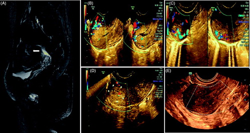
Figure 4. HIFU PRO2008 ablation therapy on placenta accreta with marked vascularity. (A,B,C) showed HIFU ablation arrays, (A) 31st slice, (B) 32nd slice, (C) 33rd slice. (D) Doppler ultrasound before HIFU, (E) Doppler ultrasound after HIFU treatment at 31st slice showed blood flow reduction.
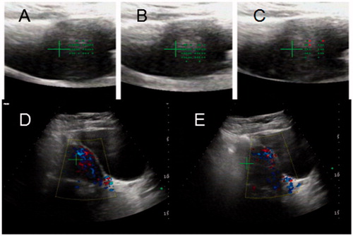
Figure 5. Placental implantation lesions and pathological findings. (A) Retained placenta accreta tissue showed tissue degeneration (HIFU); (B) Tissue degeneration and infarction under the microscope (HE × 200).
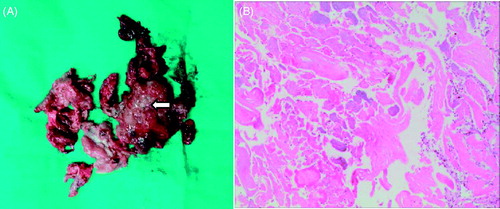
Table 2. HIFU treatment results of placenta accreta.
Table 3. Follow-up results of placenta accreta.