Figures & data
Figure 1. Preoperative and postoperative MRI. (A) Enhanced MRI image of diffused adenomyosis before treatment, which shows that the myometrium of the posterior uterine wall was thickening with a rich vascular supply. (B) Enhanced MRI image acquired for a patient with adenomyosis 1 d after HIFU ablation, which shows a non-perfused region in the lesion. (C) T2WI image obtained 6 months after HIFU ablation for a patient with adenomyosis, which shows complete resolution of the glandular lesions and that the uterus almost recovered to its normal size.
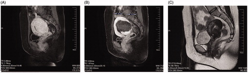
Figure 2. The VAS and VRS scores were compared before and after HIFU treatment. (A,B) The VAS (A) and VRS (B) scores decreased significantly after HIFU treatment (p < .05) for all patients diagnosed with adenomyosis. (C,D) Localized and diffused adenomyosis showed significant VAS (C) and VRS (D) score decreases after HIFU ablation (p < .05), with localized lesions achieving better therapeutic efficacy.
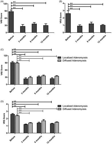
Figure 3. The menstrual volume score and hemoglobin level were compared before and after HIFU treatment. (A) The menstrual volume score decreased after HIFU treatment. (B) The hemoglobin level increased followed by HIFU ablation. (C,D) Localized and diffused adenomyosis showed significant menstrual volume decreases (C) and hemoglobin increases (D) after HIFU ablation (p < .05) and localized lesions exhibited better treatment outcomes.
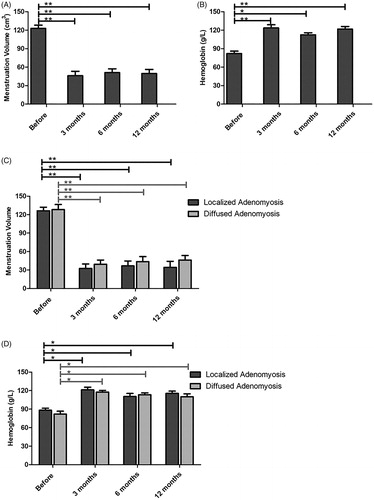
Figure 4. The uterine volume was compared before and after HIFU treatment. (A) The uterine volume was significantly reduced after HIFU ablation. (B) Localized and diffused adenomyosis showed significant uterine volume decreases after HIFU treatment, while localized lesions revealed superior clinical effectiveness.
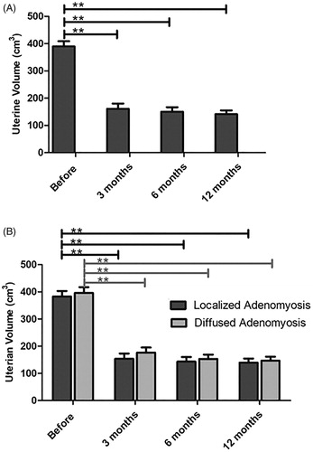
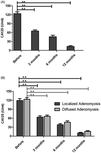
Figure 5. The serum CA-125 value was compared before and after treatment with HIFU. (A) The serum CA-125 level was significantly reduced with the combined treatment and within the normal range at 12 months after HIFU. (B) Localized and diffused adenomyosis revealed an obvious decline in CA-125 levels and better efficacy was observed for localized lesions.