Figures & data
Table 1. Ablation studies including the treatment of T1b renal masses.
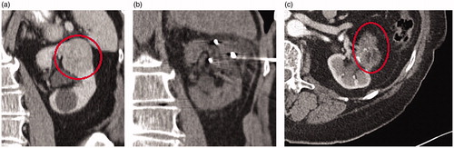
Figure 1. Seventy-two-year-old male presents with a large 7 cm right renal mass. Due to extensive medical comorbidities, he was referred for cryoablation. Preablation contrast-enhanced axial CT demonstrates a 7 cm solid exophytic mass arising from the lower pole right kidney (A). Seven cryoablation needles were placed into the mass using US/CT guidance and a 12 min freeze-5 min passive thaw-12 min refreeze was performed (B). Contrast enhanced CT at 28 months post ablation demonstrates no evidence of residual disease (C).
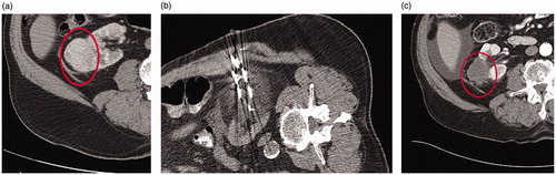
Figure 2. Forty-nine-year-old male presents with a 5 cm interpolar left renal mass. Preablation contrast-enhanced coronal CT demonstrates a heterogeneous, enhancing 5 cm mass in the interpolar left kidney (A). A biopsy during ablation revealed grade 2 clear cell renal cell carcinoma. Six cryoablation needles were used to perform a 8 min freeze- 5 min passive thaw- 10 min refreeze (B). Contrast enhanced CT five years after ablation demonstrates no evidence of recurrent RCC (C).
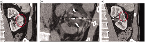
Figure 3. Eighty-year-old male presents with a 4.9 cm upper pole left renal mass. Preablation contrast enhanced coronal CT demonstrates a enhancing 4.9 cm mass in the upper pole left kidney (A). Pre ablation biopsy revealed grade 2 clear cell renal cell carcinoma. Four cryoablation needles were placed into the mass using ultrasound guidance and a 12 min freeze- 5 min passive thaw- 12 min refreeze was performed (B). Contrast enhanced coronal CT 72 months after ablation demonstrates no evidence of recurrent tumor (C).